How to use this site?

Please click on the comments to see the right option from the choices given

Dear Medicos,
This site contains a comprehensive list of medical PG entrance questions asked in various PG entrance examination throughout India like AIIMS, AIPGEE, PGI CHANDIGARH, JIPMER, CMC VELLORE .... and various state entrance exams like KERALA, TAMIL NADU, KARNATAKA, DELHI .... and also private entrances like COMEDK, MANIPAL etc...


SEARCH THE WEB

Showing posts with label ANATOMY. Show all posts

20150703

All develop from 2nd branchial arch except:

a. Buccinator
b. 7th cranial nerve
c. Anterior belly of digastric
d. Stapes


Ans c. Anterior belly

Goblet cells are present in all except?

a. Small intestine
b. Large intestine
c. Esophagus
d. Stomach

Goblet cells: glandular simple columnar epithelial cells.

     - secretes mucin, which dissolves in water to form mucus.
     - use apocrine and merocrine methods.
     - found in epithelial lining of intestinal and respiratory tracts.

: Trachea, bronchus, bronchioles
: Small intestine, Colon
: Conjunctiva in upper eyelid


Clinical significance
Goblet cell carcinoids are a class of rare tumors that form as a result of an excessive proliferation of both goblet and neuroendocrine cells. The majority of these tumors arise in the appendix and may present symptoms similar to the much more common acute appendicitis.The main treatment for localized goblet cells tumors is removal of the appendix, and sometimes removal of the right hemicolon is also performed.Disseminated tumors may require treatment with chemotherapy in addition to surgery.

Goblet cells may be an indication of metaplasia, such as in Barrett's esophagus.


Ans d. Stomach

Which layer of scalp is vascular?

a. Skin
b. Subcutaneous tissue
c. Aponeurosis
d. Loose connective tissue


Ans. b. Subcutaneous dense connective tissue

20150626

Broca's area is located in:

a. Superior frontal gyrus
b. Inferior frontal gyrus
c. Cingulate sulcus
d. Insula

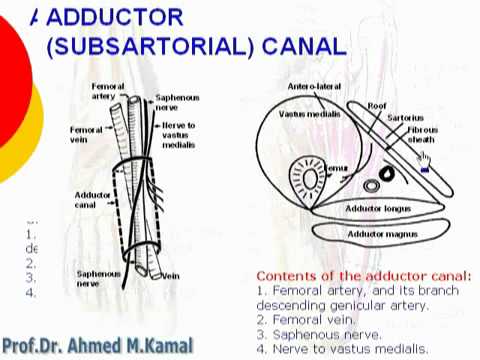
Contents of adductor canal are all except:

a. Femoral artery
b. Popliteal artery
c. Nerve to vastus medialis
d. Saphenous nerve

20150619

All veins open into coronary sinus except:

a. Great cardiac vein
b. Anterior cardiac vein
c. Middle cardiac vein
d. Left posterior ventricular vein


Ans . b. Anterior

Evertor of ankle inserted into the medial cuneiform is:


a. Peroneus longus
b. Peroneus brevis
c. Tibialis anterior
d. Tibialis posterior


20150617

Taste sensation of anterior two third of tongue is?

a. Facial nerve
b. Glossopharyngeal nerve
c. Lingual nerve
d. Vagus nerve


IMG_0851[1]
Courtesy; theninththeory.com
Ans. Lingual

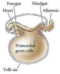
Primordial germ cell develops from:

a. Chorion
b. Amnion
c. Yolk sac
d. Cord blood


Ans. c) Yolk sac

20150616

Artery felt at anterior border of masseter against maxillary:

a. Facial artery
b. Maxillary artery
c. Posterior auricular artery
d. Superficial temporal artery


Branches external carotid artery : mnemonic
  • Some American Ladies Found Our Pyramids Most Satisfactory
  • Some Anatomists Like Freaking Out Poor Medical Students
  • Some Anatomists Like F-ing, Others Prefer S&M


Ans: a. Facial artery

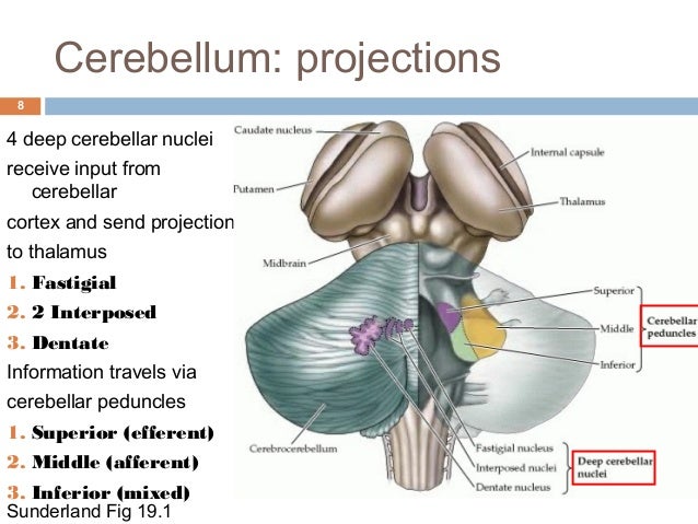
Most lateral deep cerebellar nucleus is:

a. Emboliform nucleus
b. Dentate nucleus
c. Globose nucleus
d. Fastigial nucleus

 
From lateral to medial, the four deep cerebellar nuclei are the dentate, emboliform, globose, and fastigii. Some animals, including humans, do not have distinct emboliform and globose nuclei, instead having a single, fused interposed nucleus. In animals with distinct emboliform and globose nuclei, the term interposed nucleus is often used to refer collectively to these two nuclei.

Ans: b. Dentate ( Teeth on the outside)

Cells present in cerebellum are all except:

a. Purkinje cells
b. Stellate cells
c. Pyramidal cells
d. Basket cells

20150615

Which of the following is decussated in superior medullary velum?

a. 3rd nerve
b. 4th nerve
c. 5th nerve
d. 6th nerve


Superior medullary velum

From Wikipedia, the free encyclopedia
Superior medullary velum
Gray701.png
Coronal section of the pons, at its upper part. (Ant. med. velum labeled at center top.)
Gray703.png
Anterior view of the cerebellum. (Ant. medullary velum labeled at center top.)

The superior medullary velum (anterior medullary velum, valve of Vieussens) is a thin, transparent lamina of white matter, which stretches between the superior cerebellar peduncles; on the dorsal surface of its lower half the folia and lingula are prolonged.
It forms, together with the superior cerebellar peduncle, the roof of the upper part of the fourth ventricle; it is narrow above, where it passes beneath the facial colliculi, and broader below, where it is continuous with the white substance of the superior vermis.
A slightly elevated ridge, the fraenulum veli, descends upon its upper part from between the inferior colliculi, and on either side of this the trochlear nerve emerges.
Blood is supplied by branches from the superior cerebellar artery.

The trochlear nerve is unique among the cranial nerves in several respects. It is the smallest nerve in terms of the number of axons it contains. It has the greatest intracranial length. Finally, it is the only cranial nerve that exits from the dorsal aspect of the brainstem.
"Brainstem trochlear". Licensed under CC BY-SA 3.0 via Wikipedia - https://en.wikipedia.org/wiki/File:Brainstem_trochlear.png#/media/File:Brainstem_trochlear.png

Which is a typical intercostal nerve?

a. First
b. Second
c. Third
d. Seventh


Intercostal nerves can be divided into atypical and typical intercostal nerves:
  • atypical
    • first two intercostal nerves (T1 and T2) in addition to supplying the thorax also give branches to the brachial plexus
    • lowest five intercostal nerves (T7-T11) also supply the abdomen and peritoneum
  • typical
    • the remaining intercostal nerves (T3-T6) supply only the the thoracic wall
Courtesy: Radiopaedia.org


Ans c. THIRD

Basic division of anatomical segments of liver are all except:

a. Hepatic artery
b. Hepatic vein
c. Portal vein
d. Bile duct


b. Hepatic vein

20120131

Torus aorticus

The torus aorticus is the prominent region of the right atrial septum sited superiorly and anteriorly. It is superior to the coronary sinus and anterior to the fossa ovalis. It represents the deeper and anterior surface of the posterior sinus and cusp of the aorticvalve.

.

20111116

HEPATIC HILAR AREA: THE PLATE SYSTEM

 The plate system consists of bile ducts and blood vessels
surrounded by a sheath that is continuous with Glisson’s
capsule, intrahepatically, and the hepatoduodenal
ligament, extrahepatically. This system also contains a
large number of lymphatics, nerves, and a small vascular
network. 
Couinaud  states that the bile ducts and
hepatic artery are located within the plate system, but
that the portal vein is covered with a separate sheath
of loose connective tissue, and that is why the plate
containing the extrahepatic bile duct and hepatic artery
can be easily separated from the portal vein. Three
plates are found in the hilar area: the hilar plate, the
cystic plate, and the umbilical plate

To know in detail, please check this pdf file:
http://www.springerlink.com/content/euykkaghl6c09t3k/fulltext.pdf

20111110

AIIMS May 2011- Anatomy

1. In a subclavian artery block at outer border of 1st rib, all of the following arteries help in maintaning the circulation to upper limb except:

a. Subscapular artery
b. Superior thoracic artery
c. Thyrocervical trunk.
d. Suprascapluar artery.


2. Vertical crest in fundus of the internal auditory canal is known as:

a. Bill's bar.
b. Ponticulus.
c. Cog
d. Falciform crest


3. Upper limb weight is transmitted to axial skeleton by all except:

a. Coracoacromial ligament.
b. Interclavicular ligament.
c. Costoclavicular ligament.
d. Acromioclavicular ligament.


4. Trochlear nerve- all are true except:

a. Longest intracranial course.
b. Arises from dorsum of brainstem.
c. Supply ipsilateral superior oblique muscle.
d. Enters orbit through superior orbital fissure, outside annulus of Zinn.


5. Sphincter of Oddi consists of:

a. 2
b. 3
c. 4
d. 5


6. Carpal tunnel syndrome is caused by all except;

a. Amyloidosis
b. Hypothyroidism
c. Addison's disease
d. Diabetes mellitus


7. Root of mesentery is crossed by:

a. Horizontal part of duodenum
b. Left gonadal vessels.
c. Left ureter
d. Superior mesenteric artery.


8. Neurovascular bundle in axilla is surrounded by sheath derived form:

a. Prevertebral fascia
b. Pretracheal fascia
c. Clavipectoral fascia
d. Axillary sheath.


9. On contrast radiography, which among the following is false?

a. Ileum is featureless
b. Colon has haustrations
c. Jejunum is feathery
d. Distal part of duodenum has a cap.


10. Spleen projects into the following space  of peritoneal cavity:

a. Paracolic gutter.
b. Infracolic compartment
c. Left subhepatic space
d. Greater sac.


11. All of the following muscles retract the scapula except:

a. Trapezius
b. Rhomboid major
c. Rhomboid minor
d. Levator scapulae.


12. Boundary of triangle of auscultation is not formed by:

a. Serratus anterior
b. Scapula.
c. Trapezius
d. Latissimus dorsi.


13. Sternocleidomastoid is supplied by all of the following arteries except:

a. Occipital
b. Posterior auricular
c. Thyrocervical trunk
d. Superior thyroid.


14. Axillary abscess is safely drained by which approach:

a. Medial
b. Posterior
c. Lateral
d. Floor

20111019

Papez circuit


Papez circuit is a complex circuit involved in expression of emotions. It connects the limbic system with hypothalamus and thalamus.


20111017

Neck of femur - blood supply


1. Extra capsular arterial ring:

- formed by medial and lateral circumflex femoral arteries.
- chief blood supply of head & neck of femur, formed mainly by medial circumflex.
- it gives retinacular vessels that supply 2/3 rd of head.

2. Artery of Ligamentum teres

- foveal or medial epiphyseal arery.
- branch of obturator artery.
- forms main blood supply in < 8 year.

3. Intraosseous blood supply

- via metaphysis