How to use this site?

Please click on the comments to see the right option from the choices given

Dear Medicos,
This site contains a comprehensive list of medical PG entrance questions asked in various PG entrance examination throughout India like AIIMS, AIPGEE, PGI CHANDIGARH, JIPMER, CMC VELLORE .... and various state entrance exams like KERALA, TAMIL NADU, KARNATAKA, DELHI .... and also private entrances like COMEDK, MANIPAL etc...


SEARCH THE WEB

Showing posts with label OBG. Show all posts

20120115

Kegel exercises:


Kegel exercises, also called pelvic floor exercises, help strengthen the muscles that support the uterus, bladder, and bowels. Kegel exercises also help strengthen vaginal muscles.


What are the benefits of Kegel Exercises?

Pregnant women who perform Kegel exercises often find they have an easier birth. Strengthening these muscles during pregnancy can help you develop the ability to control your muscles during labor and delivery. Toning all of these muscles will also minimize two common problems during pregnancy: bladder control and hemorrhoids.
Kegel exercises are also recommended after pregnancy to promote perineal healing, regain bladder control, and strengthen pelvic floor muscles. The best thing about Kegel exercises is that they can be done anywhere and no one knows you're doing them.

How to do Kegel Exercises

    1)There a few ways to locate your kegel muscles. You can insert a finger into the vagina and try to squeeze the muscles surrounding it. Or you practice stopping the flow of urine when urinating.( although you don't want to do this too often during urination because it can actually weaken the muscles over time and/or increase your chance of a urinary infection.)
    2) Once you have located your pelvic floor muscles, contract these muscles for 5-10 seconds, then relax, repeating 10-20 times. (Make sure to empty your bladder before doing your Kegals!)
    3) Breathe normally during the exercises, and do this at least three times a day.
    4) Try not to move your leg, buttock, or abdominal muscles during the exercises.

PERFORMING PELVIC FLOOR EXERCISES:
1. Begin by emptying your bladder.
2. Tighten the pelvic floor muscles and hold for a count of 10.
3. Relax the muscle completely for a count of 10.
4. Do 10 exercises, 3 times a day (morning, afternoon, and night).
You can do these exercises at any time and any place. Most people prefer to do the exercises while lying down or sitting in a chair. After 4 - 6 weeks, most people notice some improvement. It may take as long as 3 months to see a major change.

20111124

De Lancy levels

DeLancey describes the anatomy of vaginal vault prolapse in terms of 3 levels of support 


  • Level I involves the support of the upper vagina and cervix or the vaginal cuff (in a woman who has undergone total hysterectomy) by the cardinal-uterosacral ligament complex.
  • Level II denotes the lateral support of the mid vagina to the arcus tendineus fascia pelvis (white line).
  • Level III is represented by the fusion of tissue along the base of the urethra and the distal rectovaginal septum to the perineal body.

Ritgen manouvre

Delivery of a child's head by pressure on the perineum while controlling the speed of delivery by pressure with the other hand on the head

20110919

Wied test- Pregnancy

WIED TEST: Cytohormonal stdudy to differentiate PERIMENOPAUSE from PREGNANCY.

OUTLINE: It is tough to differentiate the cause of Amenorrhea at perimenopause between 1.Reduced ovarian function & 2.Pregnancy.

Administer:Oral Diethylstilbesterol. ( synthetic estrogen ).

Response: 1.Marked proliferation vaginal epithelium upto cornified cells.
Inference: patient is not pregnant. its due to reduced ovarian estrogen.

Response: 2.Epithelial proliferation upto intermediate cell layer.no marked proliferation response.
Inference: The patient is pregnant. 

The Lemon Sign


The lemon sign refers to the shape of the fetal skull at ultrasonography (US) when the frontal bones lose their normal convex contour and appear flattened or inwardly scalloped.
This gives the skull a shape similar to that of a lemon . The sign is seen on transverse sonograms of the fetal cranium obtained at the level of the ventricles.

The lemon sign has a strong association with spina bifida. Although the exact pathogenesis is unknown, it has been postulated that the decrease in the intraspinal pressure in neonates with spina bifida causes the brain to shift downward. This shift decreases the intracranial pressure, which is reflected onto the fetal cranium. The frontal bones are the most vulnerable to the decreased intracranial pressure and respond by flattening or scalloping inward. As the fetus matures, the lemon sign disappears because the frontal bones become stronger and are able to withstand the decreased pressure.


The lemon sign is not exclusive to spina bifida. It has been seen in a variety of conditions such as encephalocele, Dandy-Walker malformation with encephaloceles, thanatophoric dysplasia, cystic hygroma, diaphragmatic hernia, agenesis of the corpus callosum, fetal hydronephrosis, and umbilical vein varix and two-vessel cord

Pregnancy Signs


Chadwick's sign (Jacquiemer's)


Chadwick's sign is a bluish discoloration of the cervixvagina, and labia caused by the hormone estrogen which results in venous congestion. It can be observed as early as 6-8 weeks afterconception


Hegar's sign


Hegar's sign is a non-sensitive indication of pregnancy in women — its absence does not exclude pregnancy. It pertains to the features of the cervix and the uterine isthmus. It is demonstrated as softening of the uterine consistency and the possibility to palpate or compress the connection between the cervix and the fundus.
The sign is usually present from 4 to 6th week till 12 weeks of pregnancy. 


Piskacek's Sign


asymmetric enlargement of the body of the pregnant uterus as a result of its enlargement in the cornual region, usually over the site of implantation.


Piskacek's sign consists noting a palpable lateral bulge or soft prominence one of the locations where the uterine tube meets the uterus. Piskacek's sign can be noted in the seventh to eight week of gestation.


Goodell's sign


It is a significant softening of the vaginal portion of the cervix.


Ladin's sign


Ladin's sign is a clinical sign of pregnancy in which there is softening in the midline of the uterusanteriorly at the junction of the uterus and cervix. It occurs at about 6 weeks gestation.


Palmer's sign: 


uterine contractions felt during bimanual examination.


Osiander's sign


Increased pulsations felt through the lateral fornices.


Mc Donald sign


Uterus becomes flexible at uterocervical junction at 7-8 weeks.


Von Fernwald sign


Softening of fundus at 4-5 weeks


Weinberg's sign


On lateral view x- rays of pregnant abdomen, overlap of the fetal skull to the maternal

spine .

Gestational sac signs on USG

Sonographic Appearances of Normal Intrauterine G-sac:
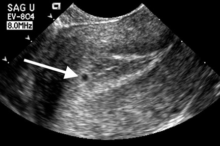
It should be possible to visualize a normal G-sac via TVS between 4.5 to 5 weeks of MGA.
The threshold level identifies the earliest we can visualize the sac :- 4 weeks + 3 days.
The discriminatory level indicates when we should always see the sac :- 5 weeks + 2 days.
The earliest appearance of gestation on TVS is referred to as Intra Decidual Sign. It is seen as a small gestational sac within within the decidua.


 Diagram of the intradecidual sign. The gestational sac (circle) does not displace or deform the central cavity complex (straight black line). The white area represents thickened decidual tissue.


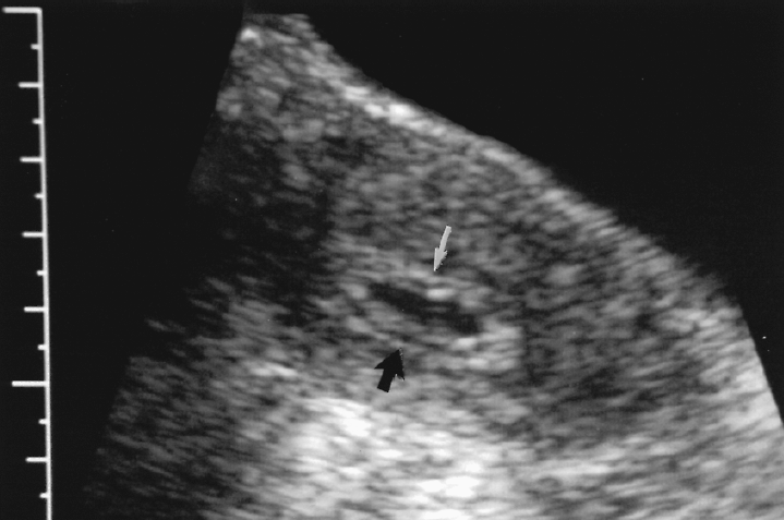
This is followed by the Double Decidual sign. It is seen as two distinct echogenic layers out of which the inner ring denotes the G-sac while the outer ring denotes the echogenic endometrial lining. This sign is helpful in differentiating it from a fluid filled decidual cast of an ectopic pregnancy.


Diagram of the double decidual sac sign. The gestational sac (circle) protrudes into and displaces the central cavity echo complex (curved line). The white area represents thickened decidual tissue.


20110918

OVARIAN TUMOURS-Histology


scanned image of page 19

Endodermal Sinus Tumour - Schiller Duval body
Schiller-Duval body is a structure seen in the endodermal sinus pattern of yolk sac tumor. It consists of a central vessel surrounded by tumor cells – the whole structure being contained in a cystic space often lined by flattened tumor cells.


Clear Cell Carcinoma - Hobnail Cells
This clear cell carcinoma shows prominent hobnail cells lining the glands. They have bulbous hyperchromatic nuclei that protrude into the gland lumen
Papillary Serous Carcinoma : Psammoma Bodies


Papillary serous carcinoma with numerous psammoma bodies infiltrating the ovarian stroma. In rare cases, psammoma bodies predominate and a present virtually in every papillae. Such tumors are termed Psammocarcinoma. They are low-grade, invasive, with small epithelial nests, and have excellent prognosis.


Granulosa Cell Tu
mor : Call-Exner bodies