How to use this site?

Please click on the comments to see the right option from the choices given

Dear Medicos,
This site contains a comprehensive list of medical PG entrance questions asked in various PG entrance examination throughout India like AIIMS, AIPGEE, PGI CHANDIGARH, JIPMER, CMC VELLORE .... and various state entrance exams like KERALA, TAMIL NADU, KARNATAKA, DELHI .... and also private entrances like COMEDK, MANIPAL etc...


SEARCH THE WEB

Showing posts with label ORTHO. Show all posts

20111124

Ponsetti method

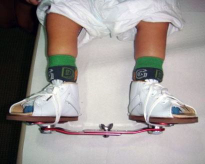
Ignacio V. Ponseti can be credited with developing a comprehensive technique for treating congenital clubfoot in the 1940s. One of the major principles of this technique is the concept that the tissues of a newborn's foot, including tendons, ligaments, joint capsules, and certain bones, will yield to gentle manipulation and casting of the feet at weekly intervals. By applying this technique to clubfeet within the first few weeks of life, most clubfeet can be successfully corrected without the need for major reconstructive surgery.


The Ponseti Technique

The corrective process utilizing the Ponseti technique can be divided into two phases:
  • the treatment phase, during which time the deformity is corrected completely, and
  • the maintenance phase, during which time a brace is utilized to prevent recurrence.
During each of these phases, attention to the details of the technique is essential to minimize the possibility of incomplete correction and recurrences.
  • The Treatment Phase
    The treatment phase should begin as early as possible, optimally within the first week of life. Gentle manipulation and casting is performed on a weekly basis. Each cast holds the foot in the corrected position, allowing it to gradually re-shape. Generally, five to six casts are required to fully correct the alignment of the foot and ankle. At the time of the final cast, the majority of infants (70% or higher) will require a percutaneous surgical procedure (with a small incision through the skin) to gain adequate lengthening of their Achilles tendon.
  • The Maintenance Phase
    The final cast remains in place for three weeks, after which the infant's foot is placed into a removable orthotic device. The orthosis is worn 23 hours per day for three months and then during the night-time for several years. Failure to use the orthosis correctly may result in recurrence of the clubfoot deformity. Good results have been demonstrated at multiple centers, and long-term results indicate that foot function is comparable with that of normal feet.

20111017

Neck of femur - blood supply


1. Extra capsular arterial ring:

- formed by medial and lateral circumflex femoral arteries.
- chief blood supply of head & neck of femur, formed mainly by medial circumflex.
- it gives retinacular vessels that supply 2/3 rd of head.

2. Artery of Ligamentum teres

- foveal or medial epiphyseal arery.
- branch of obturator artery.
- forms main blood supply in < 8 year.

3. Intraosseous blood supply

- via metaphysis

20110922

Important bone tumours


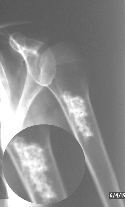
Bone infarct

"smoke up the chimney"
medullary lesion of sheet-like central lucency surrounded by sclerosis with a serpiginous border

Ewing's sarcoma


Introduction
  • malignant small round cell tumor found in young patients
  • Age & location
    • found in patients from 5-25 years of age
      • ~50% are found in the diaphysis of long bones
    • common locations include the femoral shaft, knee (distal femur and prox tibia), pelvis and proximal humerus
  • Genetics
    • 90% have t(11:22) translocation which leads to the formation of a fusion protein (EWS-FLI 1)
  • Prognosis
    • 60-70% long term survival with treatment
    • only 40% long term survival with pelvis lesions
    • only 15% long term survival if patient presents with metastasis to the lung

Symptoms
  • Presentation
    • pain often accompanied by fever
    • often mimics an infection
  • Physical exam
    • swelling and local tenderness
Imaging
  • Radiographs

    large destructive lesio
    n in the diaphysis or metaphysis with a moth-eaten appearance 
    • periosteal lifting may give "onion skin" or "sunburst" appearance
  • Bone scan
    • hot
    • required as part of workup
  • MRI often shows a large soft tissue component 
  • CT chest is important to rule out mets to lungs (very common)
Labs
  • ESR is elevated
  • WBC is elevated
  • Anemia is common
  • Bone marrow biopsy 
    • required as part of workup for Ewing's to rule out metastasis to the marrow 
Histology
  • Characteristic findings
    • sheets of monotonous small cells 
    • cells have scant cytoplasm 
    • may have pseudo-rosettes (circle of cells with necrosis in center) 
  • Immunostain
    • CD99 reactivity 
Treatment
  • Multimodality
    • multistage chemotherapy and limb salvage resection
      • preoperative chemotherapy given for 8-12 weeks followed by maintenance chemotherapy for 6-12 months after surgical resection 
    • irradiation
      • current trend towards surgical resection and away from irradiation due to high risk of irradiation-induced secondary malignancies
      • situations where radiation can be used include 
        • radiation alone for areas that cannot be resected (ie, large bulky pelvic tumors),
        • in combination with local resection if surgical margins are close

Small-round-cell tumor differential


  • if < 5 yrs. and there are small round cells think neuroblastoma or leukemia
  • if approximately 10 years think eosinophilic granuloma
  • if 10-30 yrs and there are small round cells think Ewing's
  • if >30 yrs think lymphoma
  • if > 50 yrs. and there are small round cells think myeloma

Giant cell tumour


Introduction
  • benign but aggressive tumor of unknown origin that is usually found in the epiphysis of long bones 
  • Age
    • more common in females (unlike most bone tumors)
    • ages 20-40 years (80% of giant cell tumors occur in patients older than age 20 years, with the peak incidence in the third decade of life)
      • patient age/gender on previous OITE questions: 17F, 21M, 27F, 34F, 36F, 40M 
  • Location
    • 50% around knee (distal femur or proximal tibia), most common location 
    • 10% in sacrum and vertebrae (sacrum is most common site in axial skeleton) 
    • distal radius is third most common location
    • phalanges of the hand is also a very common location
  • Malignant potential as
    • primary malignant giant cell tumor
      • metastasizes to lung in 2% 
      • hand lesions have greater chance of metastasis
    • secondary malignant giant cell tumor
      • occurs following radiation or multiple resections of giant cell tumor
  • Differential
    • If multiple lesions (~1%) than rule out hyperparathyroidism

Symptoms
  • Presentation  
    • pain referable to involved joint
Imaging
  • Radiographs
    • lytic and destructive metaphyseal lesion that often extends into the epiphysis and often borders subchondral bone 
      • may have cortical thinning with breakthrough
  • Bone scan is very hot 
  • MRI shows clear demarcation on T1 image between fatty marrow and tumor 
Histology
  • Characteristic findings
    • basic proliferating cell type is the mononucleur spindle-shaped stromal cell 
    • hallmark Giant cells are numerous 
      • nucleus of giant cell appears same as spindle-shaped stromal cells
Treatment
  • Operative
    • extensive curettage with chemical cauterization (phenol), bone grafting and cementing 
      • challenge of treatment is to remove lesion while preserving joint and providing buttress support to subchondral joint
      • aggressive exterioration of overlying cortex is required 
      • 10-30% recurrence with curettage alone verses 3% with cementation
    • hand lesion treatment is controversial
      • if no cortical breakthrough treat with curettage and cementing
      • if significant cortical breakthrough consider intercalary resection (with free fibular graft) vs. amputation
    • radiation alone for inoperable lesions only
      • leads to 15% malignant transformation

Chondrosarcoma


Introduction
  • Malignant chondrogenic lesions can occur in two forms
    • primary chondrosarcoma
      • which includes
        • low-grade, high-grade, de-differenitated chondrosarcoma
        • clear cell chondrosarcoma 
        • mesenchymal chondrosarcoma 
    • secondary chondrosarcoma
      • arises from benign cartilage lesions including 
        • osteochondroma (<1% risk of malignant transfomation)
        • multiple hereditary exostosis (1-10% risk of malignant transfomation)
        • enchondromas (1% risk of malignant transfomation)
        • Ollier's disease (25-40% risk of malignant transfomation)
        • Marfucci's (100% risk of malignant transfomation)
  • Age & location
    • typically, chondrosarcomas are found in older patients (40-75 yrs)
    • there is a slight male predominance
    • most common locations include the pelvis, proximal femur, scapula 
    • tumor location is important for diagnosis as the same histology may be diagnosed as benign in the hand but malignant if located in the long bones
  • Grade
    • 85% of chondrosarcomas are grade 1 or 2
    • 15% of chondrosarcomas are grade 3 or dedifferentiated chondrosarcoma 
      • de-differentiated chondrosarcomas are high grade lesions which develop from low grade chondroid lesions and require more aggressive treatment including adjuvant chemotherapy
  • Prognosis
    • axial and proximal extemity lesions have a more aggressive course
    • histologic grade correlates with survival
      • Grade I: 90% survival
      • Grade II: 60-70% survival
      • Grade III: 30-50% survival
      • De-differentiated chondrosarcoma: 10% survival
    • increased telomerase activity in chondrosarcoma, as determined byreverse transcriptase-polymerase chain reaction (RT-PCR), has been shown to directly correlate with the rate of reccurence 
  • Primary Chondrosarcoma sub-types
    • Clear cell chondrosarcoma 
      • malignant immature cartilaginous tumor accounting for <2% of all chondrosarcomas
      • most common in 3rd and 4th decades of life
      • commonly presents with insidious onset of pain 
      • presents as an epiphyseal lesion and can be mistaken for low-grade chondroblastoma
      • locally destructive with potential to metastasize 
    • Mesenchymal chondrosarcoma
      • chondrosarcoma variant which presents with a biphasic pattern of neoplastic cartilage with associated neoplastic small round blue cell component
      • occurs in younger patients than typical chondrosarcomas
      • may occur at several discontinuous sites at presentation and can occur in the soft tissues
      • treatment includes neo-adjuvant chemotherapy followed by wide surgical resection
Symptoms
  • Presentation
    • pain is the most common symptom
    • may present with slowly growing mass or symptoms of bowel/bladder obstruction due to mass effect in the pelvis
    • 50% of de-differentiated chondrosarcomas present with a pathologic fracture
Imaging
  • Radiographs
    • lytic or blastic lesion with reactive thickening of the cortex 
      • low-grade chondrosarcomas have a similiar appearance to enchondromas with additional cortical thickening/expansion and endosteal erosion  
      • high-grade chondrosarcomas are less well defined on plain radiographs and frequently present with cortical destruction and a soft tissue mass
    • intra-lesional "popcorn" mineralization  may be seen
      • described as rings, arcs, and stipples of mineralization
    • de-differentiated chondrosarcomas radiographically show a lower grade chondroid lesion with superimposed highly destructive area consistentwith the high grade transformed dedifferentiated chondrosarcoma 
  • MRI or CT
    • helpful to determine cortical destruction, marrow involvement, and the soft tissue involvement 
  • Bone scan
    • is usually very hot in all grades of chondrosarcoma
Histology
  • Chondrosarcoma
    • needle biopsy is not indicated for cartilage tumors due to difficulties with diagnosis
      • it is often difficult to determine malignancy based on histology alone
    • chararcteristic histology
      • low-grade chondrosarcomas show few mitotic figures with a bland histologic appearance and associated enlarged chondrocytes with plump multinucleated lacunae 
      • high-grade chondrosarcomas have a hypercellular stroma consisting of characteristic "blue-balls" of a cartilage lesion which permeate the bone trabeculae 
    • enchondromas of hand, Ollier's disease, Maffucci's disease, periosteal chondromas, and chondrosarcoma may all have similar histology
  • De-differentiated chondrosarcomas
    • characterized by a bimorphic histology
      • low grade chondroid component
      • high grade spindle cell component (similiar histology to osteosarcoma, fibrosarcoma, MFH)
Treatment
  • Operative
    • intra-lesional curettage 
      • indications
        • Grade 1 lesions 
        • treatment of grade 1 lesions located in the pelvis or axial skeleton is controversial
          • many authors recommend wide excision of all chondrosarcomas (even grade 1) if located in the pelvis
    • wide surgical excision 
      • indications
        • grade 2 or 3 lesions
        • some say grade 1 lesions in pelvis
      • historically, there is no significant role for radiation or chemotherapy in typical intramedullary chondrosarcoma 
    • wide surgical excision combined with multi-agent chemotherapy 
      • indications
        • high-grade de-differentiated chondrosarcoma
        • mesencymal chondrosarcoma

20110921

Telangiectatic osteosarcoma


Introduction
  • Occur in same location as ABC (look similar radiographically as well)

Symptoms
  • Presentation
    • 25% present with pathologic fracture
Imaging
  • Radiographs
    • lytic, destructive, and expansile lesion
  • Bone scan
    • hot
Histology
  • Characterized by
    • few cellular components (those present have malignant characteristics)
    • not as much osteoid as intramedullary osteosarcoma 
    • lakes of blood mixed with malignant cells (not in ABC) 
Treatment
  • Same as intramedullary osteosarcoma  
    • multi-agent chemotherapy and limb salvage resection
      • chemotherapy
        • preoperative chemotherapy given for 8-12 weeks followed by maintenance chemotherapy for 6-12 months after surgical resection
        • 98% necrosis with chemo is good prognostic sign
        • expression of multi-drug resistence (MDR) gene portends very poor prognosis
          • cells can pump chemo out of cell
          • present in 25% of primary lesions and 50% of metastatic lesions
      • surgical resection
        • trend towards limb salvage whenever possible
Prognosis
  • More chemosensitive but same survival as intramedullary osteosarcoma

Parosteal Osteosarcoma


Introduction
  • low grade osteosarcoma
  • Age & location
    • more common in females, age 30-40
    • previous exam question ages: 75, 74 yrs
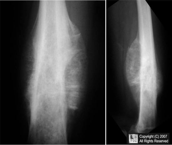
    • occurs on surface of metaphysis of long bones
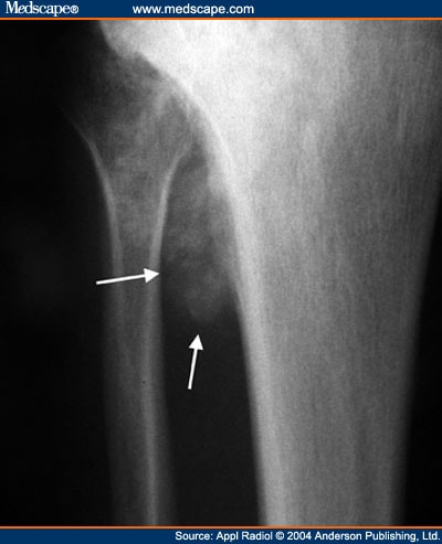
      • most common sites include posterior distal femur, proximal tibia, and proximal humerus
      • marrow invasion in 25%
Symptoms
  • Presentation
    • often presents as a painless mass
    • can limit joint motion (knee)
Imaging
  • Radiographs
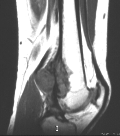
    • heavily ossified, lobulated mass arising from cortex (appears as if sticking to cortex)
  • Bone scan
    • mandatory and always hot
  • CT chest
    • mandatory to rule out pulmonary mets
  • MRI
    • mandatory to determine soft tissue involvement and skip lesions
Histology
  • Characteristic histology
    • regularly arranged normal osseous trabeculae 
    • slightly atypical spindle cells within trabeculae
    • cartilage is often present and may take the form of a cartilage cap
  • Pathologist ocassionally mistakes for fibrous dysplasia
    Treatment
    • Operative
      • wide local surgical excision
        • often curative
        • chemotherapy not indicated unless there is a high grade component
    Prognosis
    • 95% long term survival when local control has been achieved
      • dedifferentiation is a poor prognostic factor
    Groups & Differentials
    • Fibrous dysplasia (similar on histology, but xrays are different)
    • Myositis Ossificans Traumatica (juxtaposed to bone)
    • Osteochondral exostosis (shares cortex with bone)
    • Developmental defect at insertion of adductor magnus

    Osteosarcoma

    An osteosarcoma is a malignant bone forming tumour and is the second common primary bone tumour after multiple myeloma . They account for ~ 20 % of all primary bone tumours.

    Epidemiology

    Osteosarcomas can be either primary or secondary, and these have differing demographics. 
    • primary osteosarcoma : typically occurs in young patients (10 - 20 years) with 75% occurring before the age of 20  . There is a slight male predominance
    • secondary osteosarcoma : occurs in the elderly, usually secondary to malignant degeneration of Paget's disease, extensive bone infarcts or post radiotherapy for other conditions.

    Clinical presentation

    Patients usually present with bone pain, occasionally accompanied by a soft-tissue mass or swelling. At times, the first symptoms are related to pathologic fracture.
    The distribution of primary and secondary osteosarcomas is also different. 
    • primary tumours typically occur in the metaphyseal regions of long bones, and have a striking predilection for the knee, with up to 60% occurring there
    • secondary tumours on the other hand, have a much wider distribution largely mirroring the combined incidence of their underlying condition, and thus much have a higher incidence in flat bones, especially the pelvis (a favourite site of Paget's disease)

    Pathology

    Osteosarcomas can be divided into a number of sub types according to degree of differentiation, location within the bone, and histological variants 
    These sub types vary in imaging findings, demographics and biological behaviour, and include :
    Macroscopically osteosarcomas are bulky tumours where a heterogeneous cut surface demonstrates areas of haemorrhage, fibrosis and cystic degeneration. Their extension within the medullary cavity is often much more extensive than the bulky part of the tumour would suggest. Areas of bone formation are characteristic of osteosarcomas, with the degree of bone formation varying widely.
    Microscopically poorly formed trabecular bone is seen with (in the typical high grade conventional sub type) cellular pleomorphism and mitoses. Variable amounts fibrocytic and chondroblastic appearing cells may also be encountered. 
    Location
    They typically occur at the metadiaphysis of tubular bones in the appendicular skeleton. Common sites include 
    • femur : ~ 40% (especially distal femur)
    • tibia : ~ 16% (especially proximal tibia)
    • humerus : ~ 15% 
    Other less common sites include
    • fibula
    • innominate bone
    • mandible
    • maxilla
    • vertebrae  
    Markers
    Serum alkaline phosphatase (ALP) may be raised (particularly with advanced disease)

    Radiographic features

    Plain film
    Conventional radiography continues to play an important role in diagnosis. Typical appearances of conventional high grade osteosarcoma include:
    • medullary and cortical bone destruction
    • wide zone of transition, permeative or moth-eaten appearance
    • aggressive periosteal reaction
    • soft-tissue mass
    • tumour matrix ossification / calcification
      • variable:  reflects a combination of the amount of tumour bone production, calcified matrix, and osteoid
      • ill-defined "fluffy" or "cloud-like" c.f to the rings and arcs of chondroid lesions


    Chondroblastoma


    Introduction
    • A benign chondrogenic lesion 
    • Age & location
      • epiphyseal lesion in young patients (usually around 12 years of age) 
        • common locations include distal femur, proximal tibia, proximal humerus, proximal femur, and apophysis or triradiate cartilage of the pelvis
        • typically epiphyseal but may occasionally cross the physis
    • Genetics
      • may have genetic abnormalities on chromosome 5 and 8
    • Prognosis
      • 2-5% of benign chondroblasts metastasize to the lungs (similar to giant cell tumor in this respect)
    Symptoms
    • Presentation
      • pain referred to involved joint
    • Physical examination
      • exam may show mechanical symptoms with limitations of motion
    Imaging
    • Radiograph
      • well-circumscribed epiphyseal lytic lesion with thin rim of sclerotic bone that is sharply demarcated from normal medullary cavity 
        • lesions often cross physis into metaphysis
        • mineralization within the lesion may or may not be present 
    Histology
    • Classic characteristics are
      • chondroblasts arranged in "cobblestone" or "chickenwire" pattern may be present 
      • occasional multinucleated giant cells may be present 
      • loose myxoid tissue
    Treatment
    • Operative
      • extended intralesional curettage and bone grafting 
        • may do local adjuvant treatment with phenol or cryotherapy

    Enchondroma


    Introduction
    • A benign chondrogenic lesion caused by an abnormality of chondroblast function in the physis
      • chondroblasts escape the physis and proliferate in metaphysis 
      • 2nd most common benign cartilage lesion (osteochondroma is most common)
    • Age & location
      • age range is 10-60 years
      • occurs in medullary cavity in diaphysis or metaphysis
      • hand is most common location (60%)
        • presentation as pathological fractures common
      • other locations include the femur (20%) and proximal humerus (10%)
    • Include
      • solitary enchondroma
        • 1% chance of malignant transformation (chondrosarcoma)
      • Ollier's disease (multipe enchondromatosis) 
        • no genetic predisposition has been identified
        • multiple enchondromas in long bone diaphysis
        • 10-30% chance of malignant transformation into chondrosarcoma
          • however associated with high rate of both skeletal and non-skeletal malignancies
        • involved bones are dysplastic
      • Maffucci's syndrome
        • no genetic predisposition has been identified
        • multiple enchondromas and soft-tissue angiomas
        • 100% chance of malignant transformation into chondrosarcoma 
        • increased risk of visceral malignancies (astrocytoma, GI malignancy)
    Symptoms
    • Presentation
      • most are asymptomatic 
      • often present as pathologic fx of the hand but asymptomatic in foot
    • Physical exam

      • enchondroma may disrupt the growth plate and lead to shortening and angular deformities  
    Imaging
    • Radiograph
      • "pop-corn" mineralization (calcification), stippled ringsin diaphyseal or metaphyseal medullary cavity 
      • occasionally may have purly lytic appearance (especially in hand) 
    • Bone scan may be helpful and is usually minimally warm
    • MRI may show steak of cartilage or "sled runner tracks" 
    Histology
    • Classic characteristics include
      • hypocellular with bland hyaline cartilage (blue balls of cartilage)   
      • lesions in hand are more hypercellular and even display some worrisome histologic characteristics
      • small chondroid cells in lacunar spaces 
    Differential
    • Must differentiate from a bone infarct ("smoke-up-chimney" appearance)
    Treatment
    • Nonoperative
      • observation
        • treatment for most enchondromas 
        • watch with serial xrays for interval growth (obtain xrays at 3 months and 1 year from time of presentation)
    • Operative
      • curettage and bone grafting
        • indications
          • lesion that shows any change on serial xrays
          • hand fractures after fracture has first been allowed to heal with imobilization
    • Ollier's disease and Maffucci's
      • treat deformity with diaphysectomy
      • watch lesions carefully for malignant transformation