How to use this site?

Please click on the comments to see the right option from the choices given

Dear Medicos,
This site contains a comprehensive list of medical PG entrance questions asked in various PG entrance examination throughout India like AIIMS, AIPGEE, PGI CHANDIGARH, JIPMER, CMC VELLORE .... and various state entrance exams like KERALA, TAMIL NADU, KARNATAKA, DELHI .... and also private entrances like COMEDK, MANIPAL etc...


SEARCH THE WEB

Showing posts with label Harrison extracts. Show all posts

20120131

PGMCET 2012 review- Schmidt's syndrome


POLYGLANDULAR AUTOIMMUNE SYNDROME'S

When immune dysfunction affects two or more endocrine glands and other nonendocrine immune disorders are present, the polyglandular autoimmune (PGA) syndromes should be considered. The PGA syndromes are classified as two main types: 

the type I syndrome starts in childhood and is characterized by mucocutaneous candidiasis, hypoparathyroidism, and adrenal insufficiency

the type II, or Schmidt syndrome is more likely to present in adults and most commonly includes adrenal insufficiency, thyroiditis, or type 1 diabetes mellitus. Some authors have attempted to subdivide PGA II on the basis of association with some autoimmune disorders but not others (i.e., type II and type III). 

The type III syndrome is heterogeneous and may consist of autoimmune thyroid disease along with a variety of other autoimmune endocrine disorders . However, little information is gained by making this subdivision in terms of understanding pathogenesis or prevention of future endocrine complications in individual patients or in the affected families.

 Features of Polyglandular Autoimmune (Pga) Syndromes

PGA I PGA II
Epidemiology
Autosomal recessive Polygenic inheritance
Mutations in APECED gene HLA-DR3 and HLA-DR4 associated
Childhood onset Adult onset
Equal male:female ratio Female predominance
Disease Associations
Mucocutaneous candidiasis Adrenal insufficiency
Hypoparathyroidism Hypothyroidism
Adrenal insufficiency Graves' disease
Hypogonadism Type 1 diabetes
Alopecia Hypogonadism
Hypothyroidism Hypophysitis
Dental enamel hypoplasia Myasthenia gravis
Malabsorption Vitiligo
Chronic active hepatitis Alopecia
Vitiligo Pernicious anemia
Pernicious anemia Celiac disease

Abbreviation: APECED, autoimmune polyendocrinopathy-candidiasis-ectodermal dystrophy.

Polyglandular Autoimmune Syndrome Type I

PGA type I usually is recognized in the first decade of life and requires two of three components for diagnosis: mucocutaneous candidiasis, hypoparathyroidism, and adrenal insufficiency. Mucocutaneous candidiasis and hypoparathyroidism present with similar high frequency (100% and 79–96%, respectively). Adrenal insufficiency is observed in 60–72% of patients. Mineralocorticoids and glucocorticoids may be lost simultaneously or sequentially. 

PGA type 1 also is called autoimmune polyendocrinopathy-candidiasis-ectodermal dystrophy (APECED). Other endocrine defects can include gonadal failure (60% female, 14% male), hypothyroidism (5%), and destruction of the beta cells of the pancreatic islets and development of insulin-dependent (type 1) diabetes mellitus (14% lifetime risk). Additional features include hypoplasia of the dental enamel, nail dystrophy, tympanic membrane sclerosis, vitiligo, keratopathy, and gastric parietal cell dysfunction resulting in pernicious anemia (13%). Some patients develop autoimmune hepatitis (12%), malabsorption (variably attributed to intestinal lymphangiectasia, bacterial overgrowth, or hypoparathyroidism), asplenism, achalasia, and cholelithiasis . At the outset, only one organ may be involved, but the number increases with time so that patients eventually manifest two to five components of the syndrome.

Most patients initially present with oral candidiasis in childhood; it is poorly responsive to treatment  and relapses frequently. Chronic hypoparathyroidism usually occurs before adrenal insufficiency develops. More than 60% of postpubertal women develop premature hypogonadism. The endocrine components, including adrenal insufficiency and hypoparathyroidism, may not develop until the fourth decade, making continued surveillance necessary.

Type I PGA syndrome is not associated with a particular HLA type and usually is inherited as an autosomal recessive trait. It may occur sporadically. The responsible gene, designated as either APECED or AIRE, encodes a transcription factor that is expressed in thymus and lymph nodes; a variety of different mutations have been reported. The mechanism by which these mutations lead to the diverse manifestations of type I PGA is unknown.


Polyglandular Autoimmune Syndrome Type II:

PGA type II is characterized by two or more of the endocrinopathies listed above. Most often these endocrinopathies include primary adrenal insufficiency, Graves' disease or autoimmune hypothyroidism, type 1 diabetes mellitus, and primary hypogonadism. Because adrenal insufficiency is relatively rare, it is used frequently to define the presence of the syndrome. Among patients with adrenal insufficiency, type 1 diabetes mellitus coexists in 52% and autoimmune thyroid disease occurs in 69%. However, many patients with antimicrosomal and antithyroglobulin antibodies never develop abnormalities of thyroid function. Thus, increased antibody titers alone are poor predictors of future disease. Other associated conditions include hypophysitis, celiac disease (2–3%), atrophic gastritis, and pernicious anemia (13%). Vitiligo, which is caused by antibodies against the melanocyte, and alopecia are less common than in the type I syndrome. Mucocutaneous candidiasis does not occur. A few patients develop a late-onset, usually transient hypoparathyroidism caused by antibodies that compete with PTH for binding to the PTH receptor. Up to 25% of patients with myasthenia gravis and an even higher percentage who have myasthenia and a thymoma have PGA type II.

The type II syndrome is familial in nature, often transmitted as an autosomal dominant trait with incomplete penetrance. As in many of the individual autoimmune endocrinopathies, certain HL-DR3 and -DR4 alleles increase disease susceptibility; several different genes probably contribute to the expression of this syndrome.

A variety of autoantibodies are seen in PGA type II, including antibodies directed against 

(1) thyroid antigens such as thyroid peroxidase, thyroglobulin, and the thyroid-stimulating hormone (TSH) receptor; 

(2) adrenal side chain cleavage enzyme, steroid 21-hydroxylase, or ACTH receptor; and 

(3) pancreatic islet glutamic acid decarboxylase or the insulin receptor, among others. 

The roles of cytokines such as interferon and cell-mediated immunity are unclear.

Diagnosis:

The clinical manifestations of adrenal insufficiency often develop slowly, may be difficult to detect, and can be fatal if not diagnosed and treated appropriately. Thus, prospective screening should be performed routinely in all patients and family members at risk for PGA types I and II. The most effective screening test for adrenal disease is a cosyntropin stimulation test . A fasting blood glucose level can be obtained to screen for hyperglycemia. Additional screening tests should include measurements of TSH, luteinizing hormone, follicle-stimulating hormone, and, in men, testosterone levels. In families with suspected type I PGA syndrome, calcium and phosphorus levels should be measured. These screening studies should be performed every 1–2 years up to about age 50 in families with PGA type II syndrome and until about age 40 in patients with type I syndrome. Screening measurements of autoantibodies against potentially affected endocrine organs are of uncertain prognostic value. 

The differential diagnosis of PGA syndrome should include the 

-DiGeorge syndrome (hypoparathyroidism due to glandular agenesis and mucocutaneous candidiasis) 
-Kearns-Sayre syndrome (hypoparathyroidism, primary hypogonadism, type 1 diabetes mellitus, and panhypopituitarism)
-Wolfram's syndrome (congenital diabetes insipidus and diabetes mellitus)
-IPEX syndrome (immunodysregulation, polyendocrinopathy, and enteropathy, X-linked)
-congenital rubella (type 1 diabetes mellitus and hypothyroidism).


Treatment: Polyglandular Autoimmune Syndrome

With the exception of Graves' disease, the management of each of the endocrine components of the disease involves hormone replacement.  Some aspects of therapy merit special emphasis. 

Primary hypothyroidism can mask adrenal insufficiency by prolonging the half-life of cortisol; consequently, administration of thyroid hormone to a patient with unsuspected adrenal insufficiency can precipitate adrenal crisis. Thus, all patients with hypothyroidism in the context of PGA syndrome should be screened for adrenal disease and, if it is present, treated with glucocorticoids before or concurrently with thyroid hormone therapy. 

Hypoglycemia or decreasing insulin requirements in a patient with diabetes mellitus type 1 may be the earliest symptom of adrenal insufficiency. Consequently, such patients should be screened for adrenal disease. 

Treatment of mucocutaneous candidiasis with ketoconazole may induce adrenal insufficiency. This drug also may elevate liver enzymes, making the diagnosis of autoimmune hepatitis more difficult. 

Hypocalcemia in PGA type II is more commonly due to malabsorption associated with celiac disease than to hypoparathyroidism.

20111116

EVIDENCE-BASED MEDICINE (AIIMS NOV 2011 Discussion)


The "art of medicine" is defined traditionally as a practice combining medical knowledge (including scientific evidence), intuition, and judgment in the care of patients . EBM updates this construct by placing much greater emphasis on the processes by which clinicians gain knowledge of the most up-to-date and relevant clinical research to determine for themselves whether medical interventions alter the disease course and improve the length or quality of life. The meaning of practicing EBM becomes clearer through an examination of its four key steps:
  1. Formulating the management question to be answered
  2. Searching the literature and online databases for applicable research data
  3. Appraising the evidence gathered with regard to its validity and relevance
  4. Integrating this appraisal with knowledge about the unique aspects of the patient (including the patient's preferences about the possible outcomes)

Step 1 involves generating well-formulated questions that involve four or five components—PICOD: patient or population, intervention, comparator, outcome, and, sometimes, D for study design, (e.g., does routine percutaneous coronary intervention improve survival compared with initial medical management in 60-year-old men with stable angina and known CAD?) Steps 2 and 3 are the heart of EBM as it is currently used in practice and relate to the underlying fundamental principle that the strength of medical evidence supporting a therapy or strategy is hierarchical. The process of searching the world's research literature and appraising the quality and relevance of studies thus identified can be quite time-consuming and requires skills and training that most clinicians do not possess. Thus, the best starting point for most EBM searches is the identification of recent systematic overviews of the problem in question (Table 3-3).

Table 3-3 Selected Tools for Finding the Evidence in Evidence-Based Medicine
NameDescriptionWeb AddressAvailability
Evidence-Based Medicine ReviewsComprehensive electronic database that combines and integrates:
1. The Cochrane Database of Systematic Reviews
2. ACP Journal Club
3. The Database of Abstracts of Reviews of Effectiveness
www.ovid.comSubscription required. Available through medical center libraries and other institutions.
Cochrane LibraryCollection of EBM databases, including The Cochrane Database of Systematic Reviews—full text articles reviewing specific health care topics.www.cochrane.orgSubscription required. Abstracts of systematic reviews available free online. Some countries have funding to provide free access to all residents.
ACP Journal ClubCollection of summaries of original studies and systematic reviews. Published bimonthly. All data since 1991 available on Web site, updated yearly.www.acpjc.orgSubscription required.
Clinical EvidenceMonthly updated directory of concise overviews of common clinical interventions.www.clinicalevidence.comSubscription required. Free access for United Kingdom and developing countries.
MEDLINENational Library of Medicine database with citations back to 1966.www.nlm.nih.govFree via Internet.


Generally, the EBM tools listed in Table 3-3 provide access to research information in one of two forms. The first, primary research reports, is the original peer-reviewed research work that is published in medical journals. Initial access to this information in an EBM search may be gained through MEDLINE, which provides access to a huge amount of data in abstract form. However, in using MEDLINE it is often difficult to locate reports that are on point in a sea of irrelevant or unhelpful information and be reasonably certain that important reports have not been overlooked. The second form, systematic reviews, comprehensively summarizes the available evidence on a particular topic up to a certain date and provides the interpretation of the reviewer and thus is the highest level of evidence in the hierarchy. Explicit criteria are used to find all the relevant scientific research and grade its quality. The prototype for this kind of resource is the Cochrane Database of Systematic Reviews. One of the key components of a systematic review is a meta-analysis. The next two sections will review some of the major types of clinical research reports available in the literature and the process of aggregating those data into meta-analyses.

SOURCES OF EVIDENCE: CLINICAL TRIALS AND REGISTRIES

The notion of learning from observation of patients is as old as medicine itself. Over the last 50 years, physicians' understanding of how best to turn raw observation into useful evidence has evolved considerably. Case reports, personal anecdotal experience, and small single-center case series are now recognized as having severe limitations in validity and generalizability, and although they may generate hypotheses or be the first reports of adverse events, they have no role in formulating modern standards of practice. The major tools used to develop reliable evidence consist of the randomized clinical trial and the large observational registry. A registry or database typically is focused on a disease or syndrome (e.g., cancer, CAD, heart failure), a clinical procedure (e.g., bone marrow transplantation, coronary revascularization), or an administrative process (e.g., claims data used for billing and reimbursement).

By definition, in observational data, the care of the patient is not controlled by the investigator. Carefully collectedprospective observational data can achieve a level of quality approaching that of major clinical trial data. At the other end of the spectrum, data collected retrospectively (e.g., chart review) are limited in form and content to what previous observers thought was important to record, which may not serve the research question under study particularly well. Data not specifically collected for research (e.g., claims data) often have important limitations that cannot be overcome in the analysis phase of the research. Advantages of observational data include the ability to capture a broader population than is typically represented in clinical trials because of inclusion and exclusion criteria. In addition, observational data are the primary source of evidence for questions for which a randomized trial cannot or will not be performed. For example, it may be difficult or unethical to randomize patients to test diagnostic or therapeutic strategies that are unproven but widely accepted in practice. In addition, patients cannot be randomized to a sex, racial/ethnic group, socioeconomic status, or country of residence. Physicians are also not willing to randomize patients to a potentially harmful intervention, such as smoking or overeating to develop obesity.

The major difference between a well-done randomized clinical trial and a well-done prospective observational study of a particular management strategy is the lack of protection from treatment selection bias in the latter. The use of observational data to compare diagnostic or therapeutic strategies assumes that there is sufficient uncertainty in practice to ensure that similar patients will be managed differently by different physicians. In short, the analysis assumes that there is an element of randomness (in the sense of disorder rather than in the formal statistical sense) to clinical management. In such cases, statistical models attempt to adjust for important imbalances and "level the playing field" so that a fair comparison among treatment options can be made. When management is clearly not random (e.g., all eligible left main coronary artery disease patients are referred for coronary bypass surgery), the problem may be too confounded (biased) for statistical correction, and observational data may not provide reliable evidence.

In general, the use of concurrent controls is vastly preferable to that of historical controls. For example, comparison of current surgical management of left main CAD with left main CAD patients treated medically during the 1970s (the last time these patients were routinely treated with medicine alone) would be extremely misleading since the quality of "medical therapy" has made substantial improvements in the interval.

Randomized controlled clinical trials include the careful prospective design features of the best observational data studies but also include the use of random allocation of treatment. This design provides the best protection against confounding due to treatment selection bias (a major aspect of internal validity). However, the randomized trial may not have good external validity (generalizability) if the process of recruitment into the trial resulted in the exclusion of many potentially eligible subjects.

Consumers of medical evidence need to be aware that randomized trials vary widely in their quality and applicability to practice. The process of designing such a trial often involves a great many compromises. For example, trials designed to gain U.S. Food and Drug Administration (FDA) approval for an investigational drug or device have to address certain regulatory requirements that may result in a trial design different from what practicing clinicians would find useful.

META-ANALYSIS

The Greek prefix meta signifies something at a later or higher stage of development. Meta-analysis is research done on research data for the purpose of combining and summarizing the available evidence quantitatively. Although it can be used to combine nonrandomized studies, meta-analysis is used most typically to summarize all the randomized trials on a particular therapeutic problem. Ideally, unpublished trials should be identified and included to avoid publication bias (i.e., "negative" trials may not be published). Furthermore, some of the best meta-analyses obtain and analyze the raw individual patient-level data from all trials rather than working only with what is available in the published reports of each trial. Not all published meta-analyses are reliable sources of evidence on a particular problem. Their methodology must be scrutinized carefully to ensure proper study design and analysis. The results of a well-done meta-analysis are likely to be most persuasive if they include at least several large-scale, properly performed randomized trials. Although meta-analysis can help detect benefits when individual trials are inadequately powered (e.g., the benefits of streptokinase thrombolytic therapy in acute MI demonstrated by ISIS-2 in 1988 were evident by the early 1970s through meta-analysis), in cases in which the available trials are small or poorly done, meta-analysis should not be viewed as a remedy for the deficiency in primary trial data.

Meta-analyses typically focus on summary measures of relative treatment benefit, such as odds ratios or relative risks. Clinicians also should examine what absolute risk reduction (ARR) can be expected from the therapy. A useful summary metric of absolute treatment benefit is the number needed to treat (NNT) to prevent one adverse outcome event (e.g., death, stroke). NNT is simply 1/ARR

For example, if a hypothetical therapy reduced mortality rates over a 5-year follow-up by 33% (the relative treatment benefit) from 12% (control arm) to 8% (treatment arm), the absolute risk reduction would be 12% – 8% = 4% and the NNT would be 1/.04, or 25. Thus, it would be necessary to treat 25 patients for 5 years to prevent 1 death. If the hypothetical treatment was applied to a lower-risk population, say, with a 6% 5-year mortality, the 33% relative treatment benefit would reduce absolute mortality by 2% (from 6 to 4%), and the NNT for the same therapy in this lower-risk group of patients would be 50. Although not always made explicit, comparisons of NNT estimates from different studies should account for the duration of follow-up used to create each estimate

20110930

Atypical Colitides


Two atypical colitides—collagenous colitis and lymphocytic colitis—have completely normal endoscopic appearances. 
Collagenous colitis has two main histologic components: increased subepithelial collagen deposition and colitis with increased intraepithelial lymphocytes. The female to male ratio is 9:1, and most patients present in the sixth or seventh decades of life. The main symptom is chronic watery diarrhea. Treatments range from sulfasalazine or mesalamine and Lomotil to bismuth to budesonide to prednisone for refractory disease.
Lymphocytic colitis has features similar to collagenous colitis, including age at onset and clinical presentation, but it has almost equal incidence in men and women and no subepithelial collagen deposition on pathologic section. However, intraepithelial lymphocytes are increased. The frequency of celiac disease is increased in lymphocytic colitis and ranges from 9 to 27%. Celiac disease should be excluded in all patients with lymphocytic colitis, particularly if diarrhea does not respond to conventional therapy. Treatment is similar to that of collagenous colitis with the exception of a gluten-free diet for those who have celiac disease.
Diversion colitis is an inflammatory process that arises in segments of the large intestine that are excluded from the fecal stream. It usually occurs in patients with ileostomy or colostomy when a mucus fistula or a Hartmann's pouch has been created. Clinically, patients have mucus or bloody discharge from the rectum. Erythema, granularity, friability, and, in more severe cases, ulceration can be seen on endoscopy. Histopathology shows areas of active inflammation with foci of cryptitis and crypt abscesses. Crypt architecture is normal, which differentiates it from UC. It may be impossible to distinguish from CD. Short-chain fatty acid enemas may help in diversion colitis, but the definitive therapy is surgical reanastomosis.

20110919

Adrenal imaging


Generally, benign lesions are rounded and homogenous whereas most malignant lesions appear lobulated and inhomogeneous. Pheochromocytoma and adrenomyelolipoma may also exhibit lobulated and inhomogeneous features. Additional information can be obtained from CT by assessment of contrast wash-out after 15 minutes, which is >50% in benign lesions but <40% in malignant lesions, which usually have a more extensive vascularization. 


MRI also allows for the visualization of the adrenal glands with somewhat lower resolution than CT. However, as it does not involve exposure to ionizing radiation, it is preferred in children, young adults, and during pregnancy. MRI has a valuable role in the characterization of indeterminate adrenal lesions using chemical shift analysis, with malignant tumors rarely showing loss of signal on opposed-phase MRI.
Fine-needle aspiration (FNA) or CT-guided biopsy of an adrenal mass is almost never indicated. FNA of a pheochromocytoma can cause a life-threatening hypertensive crisis. FNA of an adrenocortical carcinoma violates the tumor capsule. FNA should only be considered in a patient with a history of nonadrenal malignancy and a newly detected adrenal mass. FNA should be carried out only after careful exclusion of pheochromocytoma and if the outcome will influence therapeutic management

20110915

SIADH- Syndrome of Inappropriate (Increased) ADH

Hyponatremia


CLINICAL CHARACTERISTICS
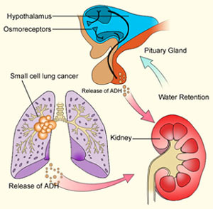
Excessive secretion or action of AVP results in the production of decreased volumes of more highly concentrated urine. If not accompanied by a commensurate reduction in fluid intake or an increase in insensible loss, the reduction in urine output results in excess water retention with expansion and dilution of all body fluids. In some patients, excessive intake results from inappropriate thirst. If the hyponatremia develops gradually or has been present for more than a few days, it may be largely asymptomatic. However, if it develops acutely, it usually is accompanied by symptoms and signs of water intoxication that may include mild headache, confusion, anorexia, nausea, vomiting, coma, and convulsions. Severe hyponatremia may be lethal. Other clinical signs and symptoms vary greatly, depending on the pathogenesis of the defect in antidiuretic function.
ETIOLOGY
Hyponatremia and impaired urinary dilution can be caused by either a primary or a secondary defect in the regulation of AVP secretion or action. The primary forms are generally referred to as the syndrome of inappropriate antidiuresis.
They have many different causes, including ectopic production of AVP by lung cancer or other neoplasms; eutopic release by various diseases or drugs; and exogenous administration of AVP, desmopressin, or large doses of oxytocin . The ectopic forms result from abnormal expression of the AVP-NPII gene by primary or metastatic malignancies. The eutopic forms occur most often in patients with acute infections or strokes but have also been associated with many other neurologic diseases and injuries. In this case, the SIAD is usually self-limited and remits spontaneously within 2–3 weeks, but about 10% of cases are chronic. The mechanisms by which these diseases disrupt osmoregulation are not known. The defect in osmoregulation can take any of four distinct forms . 
In one of the most common (reset osmostat), AVP secretion remains fully responsive to changes in plasma osmolarity/sodium but the threshold, or set point, of the osmoregulatory system is abnormally low. These patients differ from those with the other types of osmoregulatory defect in that they are able to maximally suppress plasma AVP and dilute their urine if their fluid intake is high enough to reduce their plasma osmolarity/sodium to the new set point. 
Another, smaller subgroup (10% of the total) has inappropriate antidiuresis without a demonstrable defect in the osmoregulation of plasma AVP . In some of them, all young boys, the inappropriate antidiuresis has been traced to a constitutively activating mutation of the V2 receptor gene. This unusual variant may be referred to as familial nephrogenic SIAD to distinguish it from other possible causes of the syndrome.

Causes of Syndrome of Inappropriate Antidiuresis (Siad)
Neoplasms
  Carcinomas
    Lung
    Duodenum
    Pancreas
    Ovary
    Bladder, ureter
  Other neoplasms
    Thymoma
    Mesothelioma
    Bronchial adenoma
    Carcinoid
    Gangliocytoma
    Ewing's sarcoma
Head trauma (closed and penetrating)
Infections
  Pneumonia, bacterial or viral
  Abscess, lung or brain
  Cavitation (aspergillosis)
  Tuberculosis, lung or brain
  Meningitis, bacterial or viral
  Encephalitis
  AIDS
Vascular
  Cerebrovascular occlusions, hemorrhage
  Cavernous sinus thrombosis
Genetic
X-linked recessive
(V2 receptor gene)
 
Neurologic
  Guillain-Barré syndrome
  Multiple sclerosis
  Delirium tremens
  Amyotrophic lateral sclerosis
  Hydrocephalus
  Psychosis
  Peripheral neuropathy
Congenital malformations
  Agenesis corpus callosum
  Cleft lip/palate
  Other midline defects
Metabolic
  Acute intermittent porphyria
  Pulmonary
  Asthma
  Pneumothorax
  Positive-pressure respiration
Drugs
  Vasopressin or desmopressin
  Chlorpropamide
  Oxytocin, high dose
  Vincristine
  Carbamazepine
  Nicotine
  Phenothiazines
  Cyclophosphamide
  Tricyclic antidepressants
  Monoamine oxidase inhibitors
  Serotonin reuptake inhibitors


The secondary forms of osmotically inappropriate antidiuresis also have multiple causes. They usually are subdivided into three types, depending on the nature of the abnormal stimulus and the state of extracellular fluid volume.
Type I occurs in sodium-retaining, edema-forming states such as congestive heart failure, cirrhosis, and nephrosis. It is associated with markedly excessive retention of water and sodium that is thought to be stimulated by a large reduction in "effective" blood volume caused by low cardiac output and/or redistribution of plasma from the intravascular space to the interstitial space. 
Type II occurs in sodium-depleted states such as severe gastroenteritis, diuretic abuse, and mineralocorticoid deficiency. It is due to stimulation of AVP by a large reduction in blood volume and/or pressure. In both types, the increased AVP secretion appears to be due to downward resetting of the osmostat. 
Type III is due to nonosmotic, nonhemodynamic AVP stimuli such as nausea or isolated glucocorticoid deficiency that produce a form of euvolemic hyponatremia similar to SIAD. They are differentiated because the cause of excess AVP secretion in type III can be corrected quickly and completely by treatments (antiemetics or glucocorticoids) that are not useful in SIAD.

Differential Diagnosis of Hyponatremia Based on Clinical Assessment of Extracellular Fluid Volume (Ecfv)
Clinical FindingsType I, HypervolemicType II, HypovolemicType III, EuvolemicSIAD Euvolemic
History    
CHF, cirrhosis, or nephrosisYesNoNoNo
Salt and water lossNoYesNoNo
ACTH–cortisol deficiency and/or nausea and vomitingNoNoYesNo
Physical examination    
Generalized edema, ascitesYesNoNoNo
Postural hypotensionMaybeMaybeMaybea No
Laboratory    
BUN, creatinineHigh-normalHigh-normalLow-normalLow-normal
Uric acidHigh-normalHigh-normalLow-normalLow-normal
Serum potassiumLow-normalLow-normalb Normalc Normal
Serum albuminLow-normalHigh-normalNormalNormal
Serum cortisolNormal-highNormal-highd Lowe Normal
Plasma renin activityHighHighLowf Low
Urinary sodium (meq unit of time)g LowLowh Highi Highi 

a Postural hypotension may occur in secondary (ACTH-dependent) adrenal insufficiency even though extracellular fluid volume and aldosterone are usually normal.
b Serum potassium may be high if hypovolemia is due to aldosterone deficiency.
c Serum potassium may be low if vomiting causes alkalosis.
d Serum cortisol is low if hypovolemia is due to primary adrenal insufficiency (Addison's disease).
e Serum cortisol will be normal or high if the cause is nausea and vomiting rather than secondary (ACTH-dependent) adrenal insufficiency.
f Plasma renin activity may be high if the cause is secondary (ACTH) adrenal insufficiency.
g Urinary sodium should be expressed as the rate of excretion rather than the concentration. In a hyponatremic adult, an excretion rate >25 meq/d (or 25 ueq/mg of creatinine) could be considered high.
h The rate of urinary sodium excretion may be high if the hypovolemia is due to diuretic abuse, primary adrenal insufficiency, or other causes of renal sodium wasting.
i The rate of urinary sodium excretion may be low if intake is curtailed by symptoms or treatment.
Abbreviations: ACTH, adrenocorticotropic hormone; BUN, blood urea nitrogen; CHF, congestive heart failure; SIAD, syndrome of inappropriate antidiuresis.

PATHOPHYSIOLOGY

When osmotic suppression of antidiuresis is impaired for any reason, retention of water and dilution of body fluids occur only if intake exceeds the rate of obligatory and insensible and urinary losses. The excess water intake sometimes is due to an associated defect in the osmoregulation of thirst (dipsogenic) but also can be psychogenic or iatrogenic, including IV administration of hypotonic fluids.
In SIAD, the excessive retention of water expands extracellular and intracellular volume, increases glomerular filtration and atrial natriuretic hormone, suppresses plasma renin activity, and increases urinary sodium excretion. This natriuresis reduces total body sodium, and this serves to counteract the extracellular hypervolemia but aggravates the hyponatremia. The osmotically driven increase in intracellular volume results in swelling of brain cells and increases intracranial pressure; this is probably responsible for the symptoms of acute water intoxication. Within a few days, this swelling may be counteracted by inactivation or elimination of intracellular solutes, resulting in the remission of symptoms even though the hyponatremia persists. The pathophysiology of type III (euvolemic) hyponatremia is probably similar to that of SIAD.
In type I (hypervolemic) or type II (hypovolemic) hyponatremia, the antidiuretic effect of hemodynamically induced AVP release is enhanced by decreased distal delivery of glomerular filtrate that results from increased reabsorption of sodium in proximal nephrons. If the marked reduction in urine output is not associated with a commensurate reduction in water intake or an increase in insensible loss, body fluids are expanded and diluted, resulting in hyponatremia. Unlike SIAD, however, glomerular filtration is reduced and plasma renin activity and aldosterone are elevated due to either effective hypovolemia (type I) or absolute hypovolemia (type II). Thus, urinary sodium excretion is low (unless sodium reabsorption is impaired by a diuretic), and the hyponatremia is usually accompanied by hypokalemia, azotemia, and hyperuricemia. The sodium retention is an appropriate compensatory response to severe volume and sodium depletion in type II but is inappropriate and deleterious in type I since body sodium and extracellular volume are already markedly increased, as evidenced by the presence of generalized edema.
DIFFERENTIAL DIAGNOSIS
SIAD is a diagnosis of exclusion that usually can be made from the history, physical examination, and basic laboratory data. The possibility that hyponatremia is due to an osmotically driven shift of water from the intracellular space to the extracellular space can be excluded if plasma glucose is not high enough to account for the hyponatremia [serum sodium decreases 1 meq/L for each rise in glucose of 2 mmol/L (36 mg/dL)] and/or plasma osmolarity is reduced in proportion to sodium (each decrease in serum sodium of 1 meq/L should reduce plasma osmolarity by 2 mosmol/L). The type of hypotonic hyponatremia can then be determined by standard clinical indicators of the extracellular fluid volume . If these findings are ambiguous or contradictory, measuring the rate of urinary sodium excretion or plasma renin activity may be helpful providedthat the hyponatremia is not in the recovery phase or due to a primary defect in renal conservation of sodium, diuretic abuse, or hyporeninemic hypoaldosteronism. The latter may be suspected if serum potassium is elevated instead of low as it usually is in types I and II hyponatremia. Measurements of plasma AVP are currently of no value in differentiating among the three types of hyponatremia since the abnormalities are similar. In patients who fulfill the clinical criteria for type III (euvolemic) hyponatremia, morning plasma cortisol should also be measured to exclude secondary adrenal insufficiency. If it is normal and there is no history of nausea/vomiting, the diagnosis of SIAD is confirmed and a careful search for occult lung cancer or other common causes of the syndrome should be undertaken. If an activating mutation of the V2receptor gene is suspected, plasma AVP should be measured while the hyponatremia and antidiuresis are present. If it is undetectable, DNA should be collected for analysis of the V2receptor gene.
TREATMENT: HYPONATREMIA
The management of hyponatremia differs depending not only on the type but also on the severity and duration of symptoms. In a patient with SIAD and few symptoms, the objective is to reduce body water gradually by restricting total fluid intake to less than the sum of urinary and insensible losses. Because the water derived from food (300–700 mL/d) usually approximates basal insensible losses in adults, total discretionary intake (all liquids) should be at least 500 mL less than urinary output. If achievable, this usually reduces body water and increases serum sodium by about 1–2% per day. If the symptoms or signs of water intoxication are more severe, the hyponatremia can be corrected more rapidly by supplementing the fluid restriction with IV infusion of hypertonic (3%) saline. This treatment also has the advantage of correcting the sodium deficiency that is partly responsible for the hyponatremia in SIAD and produces a solute diuresis that serves to remove some of the excess water. However, if plasma sodium is raised too rapidly or too much and the hyponatremia has been present for >24–48 hours, it also has the potential to produce central pontine myelinolysis, an acute, potentially fatal neurologic syndrome characterized by quadriparesis, ataxia, and abnormal extraocular movements. The risk of this complication can be minimized by observing several precautions: 3% saline should be infused at a rate >=0.05 mL/kg body weight per min; the effect should be monitored continuously by STAT measurements of serum sodium at least once every 2 hours; and the infusion should be stopped as soon as serum sodium increases by 12 mmol/L or to 130 mmol/L, whichever comes first. Urinary output should be monitored continuously since SIAD can remit spontaneously at any time, resulting in an acute water diuresis that greatly accelerates the rate of rise in serum sodium produced by fluid restriction and 3% saline infusion.
In chronic SIAD, the hyponatremia can be corrected by treatment with demeclocycline, 150–300 mg PO tid or gid, or fludrocortisone, 0.05–0.2 mg PO bid. The effect of the demeclocycline manifests in 7–14 days and is due to production of a reversible form of nephrogenic DI. Potential side effects include phototoxicity and azotemia. The effect of fludrocortisone also requires 1–2 weeks and is partly due to increased retention of sodium and possibly inhibition of thirst. It also increases urinary potassium excretion, which may require replacement through dietary adjustments or supplements and may induce hypertension, occasionally necessitating discontinuation of the treatment.
Nonpeptide AVP antagonists that block the antidiuretic effect of AVP have been used experimentally to treat SIAD. They produce a dose-dependent increase in urinary free-water excretion, that, if combined with a modest restriction of fluid intake, reduces body water and corrects the hyponatremia. The antagonists appear to have no adverse side effects, but, like hypertonic saline, they probably carry the risk of inducing osmotic demyelinization if the hyponatremia is corrected too rapidly. One of them, a combined V2/V1a antagonist (Conivaptan), has been approved for short-term in-hospital IV treatment of SIAD and the hyponatremia of congestive heart failure. It is a substrate and inhibitor of cytochrome P450 and should not be used in conjunction with other drugs metabolized by these pathways. Other V2 receptor antagonists are currently in phase III trials.
In type I hyponatremia, fluid restriction is also appropriate and somewhat effective if it can be maintained. However, infusion of hypertonic saline is contraindicated because it further increases total body sodium and edema and may precipitate cardiovascular decompensation. Preliminary studies with antagonists of V2 receptors indicate that they are almost as effective in type I hyponatremia as they are in SIAD.
In type II hyponatremia, the defect in AVP secretion and water balance usually can be corrected easily and quickly by stopping the loss of sodium and water and/or replacing the deficits by mouth or IV infusion of normal or hypertonic saline. As with the treatment of other forms of hyponatremia, care must be taken to ensure that plasma sodium does not increase too rapidly. Fluid restriction and administration of AVP antagonists are contraindicated in type II as they would only aggravate the underlying volume depletion and could result in hemodynamic collapse.
In euvolemic hyponatremia due to protracted nausea and vomiting or isolated glucocorticoid deficiency (type III), all abnormalities can be corrected quickly and completely by giving an antiemetic or stress doses of hydrocortisone. As with other treatments, care must be taken to ensure that serum sodium does not rise too quickly or too far.