How to use this site?

Please click on the comments to see the right option from the choices given

Dear Medicos,
This site contains a comprehensive list of medical PG entrance questions asked in various PG entrance examination throughout India like AIIMS, AIPGEE, PGI CHANDIGARH, JIPMER, CMC VELLORE .... and various state entrance exams like KERALA, TAMIL NADU, KARNATAKA, DELHI .... and also private entrances like COMEDK, MANIPAL etc...


SEARCH THE WEB

Showing posts with label CNS. Show all posts

20150616

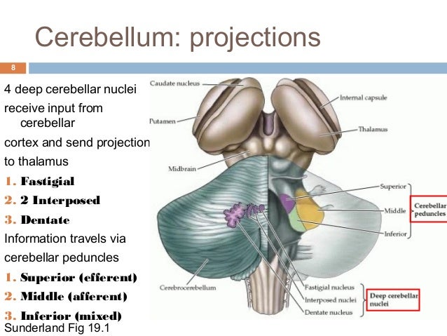
Most lateral deep cerebellar nucleus is:

a. Emboliform nucleus
b. Dentate nucleus
c. Globose nucleus
d. Fastigial nucleus

 
From lateral to medial, the four deep cerebellar nuclei are the dentateemboliformglobose, and fastigii. Some animals, including humans, do not have distinct emboliform and globose nuclei, instead having a single, fused interposed nucleus. In animals with distinct emboliform and globose nuclei, the term interposed nucleus is often used to refer collectively to these two nuclei.

Ans: b. Dentate ( Teeth on the outside)

Cells present in cerebellum are all except:

a. Purkinje cells
b. Stellate cells
c. Pyramidal cells
d. Basket cells

20150615

Which of the following is decussated in superior medullary velum?

a. 3rd nerve
b. 4th nerve
c. 5th nerve
d. 6th nerve


Superior medullary velum

From Wikipedia, the free encyclopedia
Superior medullary velum
Gray701.png
Coronal section of the pons, at its upper part. (Ant. med. velum labeled at center top.)
Gray703.png
Anterior view of the cerebellum. (Ant. medullary velum labeled at center top.)

The superior medullary velum (anterior medullary velumvalve of Vieussens) is a thin, transparent lamina of white matter, which stretches between the superior cerebellar peduncles; on the dorsal surface of its lower half the folia and lingula are prolonged.
It forms, together with the superior cerebellar peduncle, the roof of the upper part of the fourth ventricle; it is narrow above, where it passes beneath the facial colliculi, and broader below, where it is continuous with the white substance of the superior vermis.
A slightly elevated ridge, the fraenulum veli, descends upon its upper part from between the inferior colliculi, and on either side of this the trochlear nerve emerges.
Blood is supplied by branches from the superior cerebellar artery.

The trochlear nerve is unique among the cranial nerves in several respects. It is the smallest nerve in terms of the number of axons it contains. It has the greatest intracranial length. Finally, it is the only cranial nerve that exits from the dorsal aspect of the brainstem.
"Brainstem trochlear". Licensed under CC BY-SA 3.0 via Wikipedia - https://en.wikipedia.org/wiki/File:Brainstem_trochlear.png#/media/File:Brainstem_trochlear.png

20140703

Medusa head is classic for:

a. Venous malformation
b. Cavernous angioma
c. Capillary telangiectasia
d. AVM


A: a. Venous malformation.

Type of intracranial hemorrhage in battered baby:

a. Subdural
b. Extradural
c. Subarachnoid
d. Intraventricular


A: a. Subdural

Growing fracture is synonymous with:

a. Leptomeningeal cyst
b. Post-traumatic leptomeningeal cyst
c. Crista galli fracture
d. None


A: b. Post-traumatic leptomeningeal cyst

Skull fracture more often associated with epidural and subdural hematoma:

a. Linear
b. Depressed
c. Diastatic
d. All of the above


A: a. Linear

Most common location of intracranial hemorrhage with traumatic delivery is:

a. Subdural space
b. Subarachnoid space
c. Extradural space.
d. Intraventricular.


Ans. a. Subdural space