An osteosarcoma is a malignant bone forming tumour and is the second common primary bone tumour after multiple myeloma . They account for ~ 20 % of all primary bone tumours.
Epidemiology
Osteosarcomas can be either primary or secondary, and these have differing demographics.
- primary osteosarcoma : typically occurs in young patients (10 - 20 years) with 75% occurring before the age of 20 . There is a slight male predominance
- secondary osteosarcoma : occurs in the elderly, usually secondary to malignant degeneration of Paget's disease, extensive bone infarcts or post radiotherapy for other conditions.
Clinical presentation
Patients usually present with bone pain, occasionally accompanied by a soft-tissue mass or swelling. At times, the first symptoms are related to pathologic fracture.
The distribution of primary and secondary osteosarcomas is also different.
- primary tumours typically occur in the metaphyseal regions of long bones, and have a striking predilection for the knee, with up to 60% occurring there
- secondary tumours on the other hand, have a much wider distribution largely mirroring the combined incidence of their underlying condition, and thus much have a higher incidence in flat bones, especially the pelvis (a favourite site of Paget's disease)
Pathology
Osteosarcomas can be divided into a number of sub types according to degree of differentiation, location within the bone, and histological variants .
These sub types vary in imaging findings, demographics and biological behaviour, and include :
- intramedullary ~ 80%
- surface ~ 10 - 15 %
- extra skeletal ~ 5 %
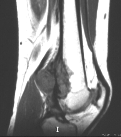
Macroscopically osteosarcomas are bulky tumours where a heterogeneous cut surface demonstrates areas of haemorrhage, fibrosis and cystic degeneration. Their extension within the medullary cavity is often much more extensive than the bulky part of the tumour would suggest. Areas of bone formation are characteristic of osteosarcomas, with the degree of bone formation varying widely.
Microscopically poorly formed trabecular bone is seen with (in the typical high grade conventional sub type) cellular pleomorphism and mitoses. Variable amounts fibrocytic and chondroblastic appearing cells may also be encountered.
Location
They typically occur at the metadiaphysis of tubular bones in the appendicular skeleton. Common sites include
- femur : ~ 40% (especially distal femur)
- tibia : ~ 16% (especially proximal tibia)
- humerus : ~ 15%
Other less common sites include
- fibula
- innominate bone
- mandible
- maxilla
- vertebrae
Markers
Serum alkaline phosphatase (ALP) may be raised (particularly with advanced disease)
Radiographic features
Plain film
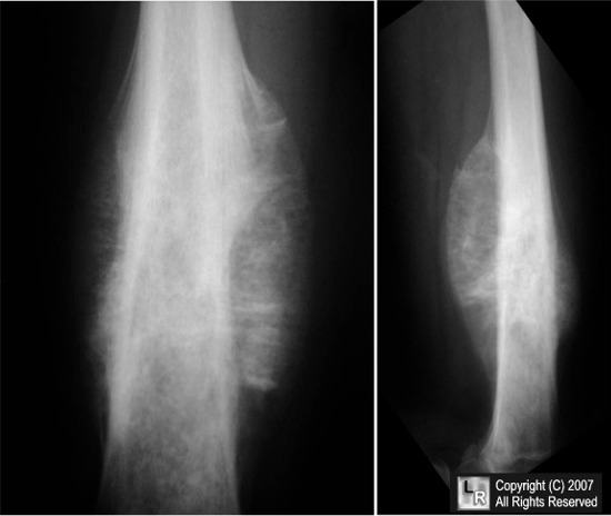
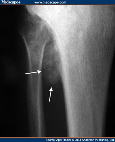
Conventional radiography continues to play an important role in diagnosis. Typical appearances of conventional high grade osteosarcoma include:

- medullary and cortical bone destruction
- wide zone of transition, permeative or moth-eaten appearance
- aggressive periosteal reaction
- soft-tissue mass
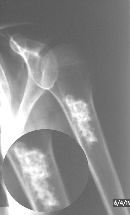
- tumour matrix ossification / calcification
- variable: reflects a combination of the amount of tumour bone production, calcified matrix, and osteoid
- ill-defined "fluffy" or "cloud-like" c.f to the rings and arcs of chondroid lesions