How to use this site?

Please click on the comments to see the right option from the choices given

Dear Medicos,
This site contains a comprehensive list of medical PG entrance questions asked in various PG entrance examination throughout India like AIIMS, AIPGEE, PGI CHANDIGARH, JIPMER, CMC VELLORE .... and various state entrance exams like KERALA, TAMIL NADU, KARNATAKA, DELHI .... and also private entrances like COMEDK, MANIPAL etc...


SEARCH THE WEB

Showing posts with label AIPGE 2011. Show all posts

20111019

Papez circuit


Papez circuit is a complex circuit involved in expression of emotions. It connects the limbic system with hypothalamus and thalamus.


20111017

Neck of femur - blood supply


1. Extra capsular arterial ring:

- formed by medial and lateral circumflex femoral arteries.
- chief blood supply of head & neck of femur, formed mainly by medial circumflex.
- it gives retinacular vessels that supply 2/3 rd of head.

2. Artery of Ligamentum teres

- foveal or medial epiphyseal arery.
- branch of obturator artery.
- forms main blood supply in < 8 year.

3. Intraosseous blood supply

- via metaphysis

Carotid artery bifurcation

Common carotid artery bifurcates at the level of upper border of thyroid cartilage, opposite the disc b/w C 3 & C 4 vertebrae.

It is usually palpated against ' Chassaignac tubercle' , ant tubercle on the transverse process of C 6 vertebra.

Heterotaxy Syndromes


Situs ambiguous also known as heterotaxy, is a rare congenital defect in which the major visceral organs are distributed abnormally within the chest and abdomen.
The normal position of the organs is known as situs solitus.
Situs inversus is a condition in which the usual positions of the organs are reversed from left to right as a mirror image of the normal condition. 
If these are the two extreme positions on a continuum of asymmetric thoracic and abdominal organ formation, situs ambiguus covers everything in between.


Situs ambiguus or heterotaxy syndrome refers
to malposition and dysmorphism of thoracic and
abdominal organs associated with indeterminate
atrial arrangement and vascular anomalies.
Azygos or hemiazygos continuation of the IVC with
absence of the hepatic segment is the most frequent associated anomaly.


Right Isomerism or Asplenia or Ivemark's syndrome:

In this condition, bilateral right sidedness occurs. These patients have bilateral right atria, a centrally located liver , an absent spleen and both lungs have 3 lobes. The descending aorta and inferior vena cava are on the same side of the spleen.

Left Isomerism or Polysplenia syndrome:


Here, bilateral left sidedness occurs. These patients have bilateral left atria and multiple spleens, and both lungs have 2 lobes. Interruption of the inferior vena cava with azygous or hemiazygous continuation is often present.

Prostatic urethra

It is the widest and most dilatable part of the male urethra.
On cross-section, it is concavo-convex in shape.

Pancreas- posterior relations

Head is related to:

1. Inferior vena cava.
2. Terminal part of renal veins.
3. Right crus of diaphragm.
4. Common bile duct.

Uncinate process :     Aorta

Neck :          Termination of superior mesenteric vein
                      beginning of portal vein.

Body:

1. Aorta and origin of SMA.
2. Left crus of diaphragm.
3. Splenic vein.
4. Left kidney.
5. Left adrenal gland.
6. Left renal vessels.


Diaphragm- development

Diaphragm develops from the following components:

1. Septum transversum
2. Pleuroperitoneal membranes.
3. Ventral and dorsal mesenteries of esophagus.
4. Mesoderm of body wall.
5. Cervical myotomes.


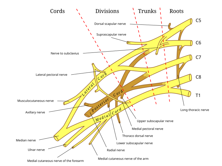
Brachial Plexus

Branches from Trunk:

1. Suprascapular Nerve ( C5, 6 )
2. N. to subclavius    ( C 5, 6 )

Branches from Roots:

1. Long thoracic nerve ( C 5, 6, 7)
2. Dorsal scapular nerve ( C 5)


Trigeminal nerve- branches


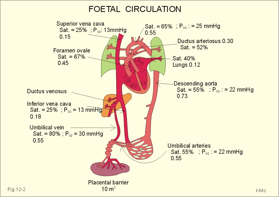
Foetal circulation

Percent saturation of foetal blood vessels

Foramen magnum


The structures passing through the foramen magnum are:

Anterior Part:      1. Apical ligament of dense.
                            2. Vertical band of cruciate ligament
                            3. Membrana tectoria

Subarachnoid Space:

1. Vertebral artery
2. Ant. Spinal artery
3. Post. Spinal artery
4. Spinal accessory nerve.
5. Sympathetic plexus around vertebral arteries.

Posterior part:  1. Lower part of medulla.
                         2. Meninges.