a. 3rd nerve
b. 4th nerve
c. 5th nerve
d. 6th nerve
b. 4th nerve
c. 5th nerve
d. 6th nerve
Superior medullary velum
From Wikipedia, the free encyclopedia
| Superior medullary velum | |
|---|---|

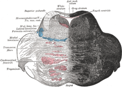
Coronal section of the pons, at its upper part. (Ant. med. velum labeled at center top.)
| |

Anterior view of the cerebellum. (Ant. medullary velum labeled at center top.)
| |
The superior medullary velum (anterior medullary velum, valve of Vieussens) is a thin, transparent lamina of white matter, which stretches between the superior cerebellar peduncles; on the dorsal surface of its lower half the folia and lingula are prolonged.
It forms, together with the superior cerebellar peduncle, the roof of the upper part of the fourth ventricle; it is narrow above, where it passes beneath the facial colliculi, and broader below, where it is continuous with the white substance of the superior vermis.
A slightly elevated ridge, the fraenulum veli, descends upon its upper part from between the inferior colliculi, and on either side of this the trochlear nerve emerges.
Blood is supplied by branches from the superior cerebellar artery.
The trochlear nerve is unique among the cranial nerves in several respects. It is the smallest nerve in terms of the number of axons it contains. It has the greatest intracranial length. Finally, it is the only cranial nerve that exits from the dorsal aspect of the brainstem.
 |
| "Brainstem trochlear". Licensed under CC BY-SA 3.0 via Wikipedia - https://en.wikipedia.org/wiki/File:Brainstem_trochlear.png#/media/File:Brainstem_trochlear.png |
No comments:
Post a Comment