How to use this site?

Please click on the comments to see the right option from the choices given

Dear Medicos,
This site contains a comprehensive list of medical PG entrance questions asked in various PG entrance examination throughout India like AIIMS, AIPGEE, PGI CHANDIGARH, JIPMER, CMC VELLORE .... and various state entrance exams like KERALA, TAMIL NADU, KARNATAKA, DELHI .... and also private entrances like COMEDK, MANIPAL etc...


SEARCH THE WEB

20111024

Hyperkinetic Movement Disorders


TremorRhythmic oscillation of a body part due to intermittent muscle contractions
DystoniaInvoluntary patterned sustained or repeated muscle contractions often associated with twisting movements and abnormal posture.
AthetosisSlow, distal, writhing, involuntary movements with a propensity to affect the arms and hands
ChoreaRapid, semipurposeful, graceful, dance-like nonpatterned involuntary movements involving distal or proximal muscle groups
MyoclonusSudden, brief (<100 ms), jerk-like, arrhythmic muscle twitches
TicBrief, repeated, stereotyped muscle contractions that are often suppressible. Can be simple and involve a single muscle group or complex and affect a range of motor activities

Guide to NSAID Therapy


 No/Low NSAID GI RiskNSAID GI Risk
No CV risk (no aspirin)Traditional NSAIDCoxib or 
  Traditional NSAID + PPI or misoprostol
  Consider non-NSAID therapy
CV risk (consider aspirin)Traditional NSAID + PPI or misoprostol if GI risk warrants gastroprotectionA gastroprotective agent must be added if a traditional NSAID is prescribed
 Consider non-NSAID therapyConsider non-NSAID therapy

20111020

Colorectal Cancer Screening Strategies


 Choices/Recommendations Comments 
Average-Risk Patients 
Asymptomatic individuals > 50 years of age (>45 years of age for African Americans)Colonoscopy every 10 years*
 
Preferred cancer prevention strategy
 Annual fecal immunochemical test (FIT) for occult bleeding, fecal DNA testing every 3 yearsCancer detection strategy; fails to detect many polyps and some cancers
 CT colonography every 5 yearsEvolving technology 
 Flexible sigmoidoscopy every 5 yearsFails to detect proximal colon polyps and cancers
 Double-contrast barium enema every 5 yearsLess sensitive than colonoscopy or CT colonography, misses some rectosigmoid polyps and cancers
Personal History of Polyps or Colorectal Cancer 
1 or 2 small (<1 cm) adenomas with low-grade dysplasiaRepeat colonoscopy in 5 yearsAssuming complete polyp resection
3 to 9 adenomas, or any adenoma >1 cm or containing high-grade dysplasia or villus featuresRepeat colonoscopy in 3 years; subsequent colonoscopy based on findingsAssuming complete polyp resection
>10 adenomasColonoscopy in <3 years based on clinical judgmentConsider evaluation for FAP or HNPCC; see recommendations below
Piecemeal removal of a sessile polypExam in 2 to 6 months to verify complete removal 
Small (<1 cm) hyperplastic polyps of sigmoid and rectumColonoscopy in 10 years 
>2 serrated polyps, or any serrated or hyperplastic polyp >1 cmRepeat colonoscopy in 3 years 
Incompletely removed serrated polyp >1 cmExam in 2 to 6 months to verify complete removal 
Colon cancerEvaluate entire colon around the time of resection, then repeat colonoscopy in 3 years 
Inflammatory Bowel Disease 
Long-standing (>8 years) ulcerative colitis or Crohn's colitis, or left-sided ulcerative colitis of >15 years' durationColonoscopy with biopsies every 1 to 3 years 
Family History of Polyps or Colorectal Cancer 
First-degree relatives with only small tubular adenomasSame as average risk 
Single first-degree relative with CRC or advanced adenoma at age >60 yearsSame as average risk 
Single first-degree relative with CRC or advanced adenoma at age <60 years, OR two first-degree relatives with CRC or advanced adenomas at any ageColonoscopy every 5 years beginning at age 40 years or 10 years younger than age at diagnosis of the youngest affected relative 
FAPSigmoidoscopy or colonoscopy annually, beginning at age 10–12 yearsConsider genetic counseling and testing
HNPCCColonoscopy every 2 years beginning at age 20–25 years until age 40, then annually thereafterConsider histologic evaluation for microsatellite instability in tumor specimens of patients who meet Bethesda criteria; consider genetic counseling and testing

20111019

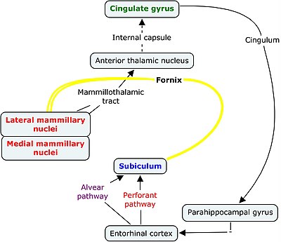
Papez circuit


Papez circuit is a complex circuit involved in expression of emotions. It connects the limbic system with hypothalamus and thalamus.


20111017

Neck of femur - blood supply


1. Extra capsular arterial ring:

- formed by medial and lateral circumflex femoral arteries.
- chief blood supply of head & neck of femur, formed mainly by medial circumflex.
- it gives retinacular vessels that supply 2/3 rd of head.

2. Artery of Ligamentum teres

- foveal or medial epiphyseal arery.
- branch of obturator artery.
- forms main blood supply in < 8 year.

3. Intraosseous blood supply

- via metaphysis

Carotid artery bifurcation

Common carotid artery bifurcates at the level of upper border of thyroid cartilage, opposite the disc b/w C 3 & C 4 vertebrae.

It is usually palpated against ' Chassaignac tubercle' , ant tubercle on the transverse process of C 6 vertebra.

Heterotaxy Syndromes


Situs ambiguous also known as heterotaxy, is a rare congenital defect in which the major visceral organs are distributed abnormally within the chest and abdomen.
The normal position of the organs is known as situs solitus.
Situs inversus is a condition in which the usual positions of the organs are reversed from left to right as a mirror image of the normal condition. 
If these are the two extreme positions on a continuum of asymmetric thoracic and abdominal organ formation, situs ambiguus covers everything in between.


Situs ambiguus or heterotaxy syndrome refers
to malposition and dysmorphism of thoracic and
abdominal organs associated with indeterminate
atrial arrangement and vascular anomalies.
Azygos or hemiazygos continuation of the IVC with
absence of the hepatic segment is the most frequent associated anomaly.


Right Isomerism or Asplenia or Ivemark's syndrome:

In this condition, bilateral right sidedness occurs. These patients have bilateral right atria, a centrally located liver , an absent spleen and both lungs have 3 lobes. The descending aorta and inferior vena cava are on the same side of the spleen.

Left Isomerism or Polysplenia syndrome:


Here, bilateral left sidedness occurs. These patients have bilateral left atria and multiple spleens, and both lungs have 2 lobes. Interruption of the inferior vena cava with azygous or hemiazygous continuation is often present.

Prostatic urethra

It is the widest and most dilatable part of the male urethra.
On cross-section, it is concavo-convex in shape.

Pancreas- posterior relations

Head is related to:

1. Inferior vena cava.
2. Terminal part of renal veins.
3. Right crus of diaphragm.
4. Common bile duct.

Uncinate process :     Aorta

Neck :          Termination of superior mesenteric vein
                      beginning of portal vein.

Body:

1. Aorta and origin of SMA.
2. Left crus of diaphragm.
3. Splenic vein.
4. Left kidney.
5. Left adrenal gland.
6. Left renal vessels.


Diaphragm- development

Diaphragm develops from the following components:

1. Septum transversum
2. Pleuroperitoneal membranes.
3. Ventral and dorsal mesenteries of esophagus.
4. Mesoderm of body wall.
5. Cervical myotomes.


Brachial Plexus

Branches from Trunk:

1. Suprascapular Nerve ( C5, 6 )
2. N. to subclavius    ( C 5, 6 )

Branches from Roots:

1. Long thoracic nerve ( C 5, 6, 7)
2. Dorsal scapular nerve ( C 5)


Trigeminal nerve- branches


Foetal circulation

Percent saturation of foetal blood vessels

Vertebral Artery- Parts


Vertebral artery is divided into the following four parts:

V 1 : Origin to transverse process of C6
V 2 : Thru f. transversaria of upper 6 cervical vertebrae.
V 3 : Sub- occipital triangle.
V 4 : Passes thru foramen magnum, pierces dura and enters subarachnoid space. 


Divisions of vertebral artery

Foramen magnum


The structures passing through the foramen magnum are:

Anterior Part:      1. Apical ligament of dense.
                            2. Vertical band of cruciate ligament
                            3. Membrana tectoria

Subarachnoid Space:

1. Vertebral artery
2. Ant. Spinal artery
3. Post. Spinal artery
4. Spinal accessory nerve.
5. Sympathetic plexus around vertebral arteries.

Posterior part:  1. Lower part of medulla.
                         2. Meninges.


Nerve supply - lower limb


Testosterone production

20111007

malnutrition- indicators

Indicators of malnutrition


Stunting - low height for age = chronic

Wasting - low weight for height = acute


Underweight - low weight for age = both acute and chronic malnutrition
"

'via Blog this'

The Parasympathetic Supply of the Head

Number 10 The Parasympathetic Supply of the Head:

Animated lectures

20111003

Neural Crest Derivatives


Connective tissue and bones of the face and skull
Cranial nerve ganglia
C cells of the thyroid gland
Conotruncal septum in the heart

Odontoblasts
Dermis in face and neck

Spinal (dorsal root) ganglia
Sympathetic chain and preaortic ganglia
Parasympathetic ganglia of the gastrointestinal tract
Adrenal medulla

Schwann cells
Glial cells
Arachnoid and pia mater (leptomeninges)
Melanocytes

Cavernous Sinus

Dural venous sinuses