How to use this site?

Please click on the comments to see the right option from the choices given

Dear Medicos,
This site contains a comprehensive list of medical PG entrance questions asked in various PG entrance examination throughout India like AIIMS, AIPGEE, PGI CHANDIGARH, JIPMER, CMC VELLORE .... and various state entrance exams like KERALA, TAMIL NADU, KARNATAKA, DELHI .... and also private entrances like COMEDK, MANIPAL etc...


SEARCH THE WEB

20110930

Atypical Colitides


Two atypical colitides—collagenous colitis and lymphocytic colitis—have completely normal endoscopic appearances. 
Collagenous colitis has two main histologic components: increased subepithelial collagen deposition and colitis with increased intraepithelial lymphocytes. The female to male ratio is 9:1, and most patients present in the sixth or seventh decades of life. The main symptom is chronic watery diarrhea. Treatments range from sulfasalazine or mesalamine and Lomotil to bismuth to budesonide to prednisone for refractory disease.
Lymphocytic colitis has features similar to collagenous colitis, including age at onset and clinical presentation, but it has almost equal incidence in men and women and no subepithelial collagen deposition on pathologic section. However, intraepithelial lymphocytes are increased. The frequency of celiac disease is increased in lymphocytic colitis and ranges from 9 to 27%. Celiac disease should be excluded in all patients with lymphocytic colitis, particularly if diarrhea does not respond to conventional therapy. Treatment is similar to that of collagenous colitis with the exception of a gluten-free diet for those who have celiac disease.
Diversion colitis is an inflammatory process that arises in segments of the large intestine that are excluded from the fecal stream. It usually occurs in patients with ileostomy or colostomy when a mucus fistula or a Hartmann's pouch has been created. Clinically, patients have mucus or bloody discharge from the rectum. Erythema, granularity, friability, and, in more severe cases, ulceration can be seen on endoscopy. Histopathology shows areas of active inflammation with foci of cryptitis and crypt abscesses. Crypt architecture is normal, which differentiates it from UC. It may be impossible to distinguish from CD. Short-chain fatty acid enemas may help in diversion colitis, but the definitive therapy is surgical reanastomosis.

Differences - Ulcerative colitis & Crohn's Disease


                                                                                          ULCERATIVE                   CROHN'S
                                                                                              COLITIS                          DISEASE

Clinical
                                                                                   
Gross blood in stoolYesOccasionally
MucusYesOccasionally
Systemic symptomsOccasionallyFrequently
PainOccasionallyFrequently
Abdominal massRarelyYes
Significant perineal diseaseNoFrequently
FistulasNoYes
Small intestinal obstructionNoFrequently
Colonic obstructionRarelyFrequently
Response to antibioticsNoYes
Recurrence after surgeryNoYes
ANCA-positiveFrequentlyRarely
ASCA-positiveRarelyFrequently
Endoscopic 
Rectal sparingRarelyFrequently
Continuous diseaseYesOccasionally
"Cobblestoning"NoYes
Granuloma on biopsyNoOccasionally
Radiographic 
Small bowel significantly abnormalNoYes
Abnormal terminal ileumNoYes
Segmental colitisNoYes
Asymmetric colitisNoYes
StrictureOccasionallyFrequently

Primary Genetic Disorders Associated with Ibd


NameGenetic AssociationPhenotype
Turner's syndromeLoss of part or all of X chromosomeAssociated with UC and colonic CD
Hermansky-PudlakAutosomal recessive chromosome 10q23Granulomatous colitis, oculocutaneous albinism, platelet dysfunction, pulmonary fibrosis
Wiskott-Aldrich syndrome (WAS)X-linked recessive disorder, loss of WAS protein functionColitis, immunodeficiency, severely dysfunctional platelets, and thrombocytopenia
Glycogen Storage diseaseDeficiency of the glucose-6-phosphate transport protein type B1Granulomatous colitis, presents in infancy with hypoglycemia, growth failure, hepatomegaly, and neutropenia
Immune dysregulation polyendocrinopathy, enteropathy X-linked (IPEX)Loss of FoxP3 transcription factor and T regulatory cell functionUC-like autoimmune enteropathy, with endocrinopathy (neonatal type 1 diabetes or thyroiditis), dermatitis
Early onset IBDDeficient IL-10 receptor functionSevere, refractory IBD in early life

Epidemiology of IBD


 Ulcerative ColitisCrohn's Disease
Incidence (North America) per person-years2.2–14.3:100,0003.1–14.6:100,000
Age of onset15–30 & 60–8015–30 & 60–80
EthnicityJewish > non-Jewish white > African American > Hispanic > Asian
Male/female ratio1:11.1–1.8:1
SmokingMay prevent diseaseMay cause disease
Oral contraceptivesNo increased riskOdds ratio 1.4
AppendectomyProtectiveNot protective
Monozygotic twins6% concordance58% concordance
Dizygotic twins0% concordance4% concordance

20110924

Amyand's hernia


Amyand's hernia is a rare form of inguinal hernia (less than 1%) which occurs when the appendix is included in the hernial sac and becomes incarcerated. The condition is an eponymous disease named after an English surgeon, Claudius Amyand , who performed the first successfulappendectomy in 1735.
Amyand's hernia is commonly misdiagnosed as an ordinary incarcerated hernia. Symptoms mimicking appendicitis may occur. Treatment consists of a combination of appendectomy and hernia repair.

20110923

Arcade of frohse

The most superior part of the superficial layer of the supinator muscle is named as the arcade of Frohse (AF). The deep branch of the radial nerve runs under this arch. The AF is reported to be the most common structure causing entrapment neuropathy of the deep branch of the radial nerve(PIN syndrome).

Osborne's ligament ( Band of Osborne )

roof of cubital tunnel is formed by aponeurosic attachment of 2 heads of FCU, which spans in arcade like manner from medial 
                epicondyle of humerus to the olecranon process of the ulna (also known as Osborne's ligament);
                - cubital tunnel begins where the ulnar nerve passes beneath Osborne's ligament;

Arcade of Struthers


    - in middle of the arm, ulnar nerve pierces medial intermuscular septum and descends in front of the medial head of the triceps;

    - in 70-80% of individuals, nerve passes under arcade of Struthers;
    - the arcade is a thin aponeurotic band extending from medial head of triceps to the medial intermuscular septum;
           - it lies approximately 8 cm proximal to the medial epicondyle;
    - the arcade may appear as medial triceps muscle fibers crossing superficial to the ulnar nerve;
    - arcade is not site for entrapment under ordinary circumstances, but it may become point of kinking if anterior transposition of ulnar nerve is performed;
           - following transposition procedures, the surgeon should release the arcade if it appears that the nerve is under tension;

20110922

Important bone tumours


Bone infarct

"smoke up the chimney"
medullary lesion of sheet-like central lucency surrounded by sclerosis with a serpiginous border

Ewing's sarcoma


Introduction
  • malignant small round cell tumor found in young patients
  • Age & location
    • found in patients from 5-25 years of age
      • ~50% are found in the diaphysis of long bones
    • common locations include the femoral shaft, knee (distal femur and prox tibia), pelvis and proximal humerus
  • Genetics
    • 90% have t(11:22) translocation which leads to the formation of a fusion protein (EWS-FLI 1)
  • Prognosis
    • 60-70% long term survival with treatment
    • only 40% long term survival with pelvis lesions
    • only 15% long term survival if patient presents with metastasis to the lung

Symptoms
  • Presentation
    • pain often accompanied by fever
    • often mimics an infection
  • Physical exam
    • swelling and local tenderness
Imaging
  • Radiographs

    large destructive lesio
    n in the diaphysis or metaphysis with a moth-eaten appearance 
    • periosteal lifting may give "onion skin" or "sunburst" appearance
  • Bone scan
    • hot
    • required as part of workup
  • MRI often shows a large soft tissue component 
  • CT chest is important to rule out mets to lungs (very common)
Labs
  • ESR is elevated
  • WBC is elevated
  • Anemia is common
  • Bone marrow biopsy 
    • required as part of workup for Ewing's to rule out metastasis to the marrow 
Histology
  • Characteristic findings
    • sheets of monotonous small cells 
    • cells have scant cytoplasm 
    • may have pseudo-rosettes (circle of cells with necrosis in center) 
  • Immunostain
    • CD99 reactivity 
Treatment
  • Multimodality
    • multistage chemotherapy and limb salvage resection
      • preoperative chemotherapy given for 8-12 weeks followed by maintenance chemotherapy for 6-12 months after surgical resection 
    • irradiation
      • current trend towards surgical resection and away from irradiation due to high risk of irradiation-induced secondary malignancies
      • situations where radiation can be used include 
        • radiation alone for areas that cannot be resected (ie, large bulky pelvic tumors),
        • in combination with local resection if surgical margins are close

Small-round-cell tumor differential


  • if < 5 yrs. and there are small round cells think neuroblastoma or leukemia
  • if approximately 10 years think eosinophilic granuloma
  • if 10-30 yrs and there are small round cells think Ewing's
  • if >30 yrs think lymphoma
  • if > 50 yrs. and there are small round cells think myeloma

Giant cell tumour


Introduction
  • benign but aggressive tumor of unknown origin that is usually found in the epiphysis of long bones 
  • Age
    • more common in females (unlike most bone tumors)
    • ages 20-40 years (80% of giant cell tumors occur in patients older than age 20 years, with the peak incidence in the third decade of life)
      • patient age/gender on previous OITE questions: 17F, 21M, 27F, 34F, 36F, 40M 
  • Location
    • 50% around knee (distal femur or proximal tibia), most common location 
    • 10% in sacrum and vertebrae (sacrum is most common site in axial skeleton) 
    • distal radius is third most common location
    • phalanges of the hand is also a very common location
  • Malignant potential as
    • primary malignant giant cell tumor
      • metastasizes to lung in 2% 
      • hand lesions have greater chance of metastasis
    • secondary malignant giant cell tumor
      • occurs following radiation or multiple resections of giant cell tumor
  • Differential
    • If multiple lesions (~1%) than rule out hyperparathyroidism

Symptoms
  • Presentation  
    • pain referable to involved joint
Imaging
  • Radiographs
    • lytic and destructive metaphyseal lesion that often extends into the epiphysis and often borders subchondral bone 
      • may have cortical thinning with breakthrough
  • Bone scan is very hot 
  • MRI shows clear demarcation on T1 image between fatty marrow and tumor 
Histology
  • Characteristic findings
    • basic proliferating cell type is the mononucleur spindle-shaped stromal cell 
    • hallmark Giant cells are numerous 
      • nucleus of giant cell appears same as spindle-shaped stromal cells
Treatment
  • Operative
    • extensive curettage with chemical cauterization (phenol), bone grafting and cementing 
      • challenge of treatment is to remove lesion while preserving joint and providing buttress support to subchondral joint
      • aggressive exterioration of overlying cortex is required 
      • 10-30% recurrence with curettage alone verses 3% with cementation
    • hand lesion treatment is controversial
      • if no cortical breakthrough treat with curettage and cementing
      • if significant cortical breakthrough consider intercalary resection (with free fibular graft) vs. amputation
    • radiation alone for inoperable lesions only
      • leads to 15% malignant transformation

Chondrosarcoma


Introduction
  • Malignant chondrogenic lesions can occur in two forms
    • primary chondrosarcoma
      • which includes
        • low-grade, high-grade, de-differenitated chondrosarcoma
        • clear cell chondrosarcoma 
        • mesenchymal chondrosarcoma 
    • secondary chondrosarcoma
      • arises from benign cartilage lesions including 
        • osteochondroma (<1% risk of malignant transfomation)
        • multiple hereditary exostosis (1-10% risk of malignant transfomation)
        • enchondromas (1% risk of malignant transfomation)
        • Ollier's disease (25-40% risk of malignant transfomation)
        • Marfucci's (100% risk of malignant transfomation)
  • Age & location
    • typically, chondrosarcomas are found in older patients (40-75 yrs)
    • there is a slight male predominance
    • most common locations include the pelvis, proximal femur, scapula 
    • tumor location is important for diagnosis as the same histology may be diagnosed as benign in the hand but malignant if located in the long bones
  • Grade
    • 85% of chondrosarcomas are grade 1 or 2
    • 15% of chondrosarcomas are grade 3 or dedifferentiated chondrosarcoma 
      • de-differentiated chondrosarcomas are high grade lesions which develop from low grade chondroid lesions and require more aggressive treatment including adjuvant chemotherapy
  • Prognosis
    • axial and proximal extemity lesions have a more aggressive course
    • histologic grade correlates with survival
      • Grade I: 90% survival
      • Grade II: 60-70% survival
      • Grade III: 30-50% survival
      • De-differentiated chondrosarcoma: 10% survival
    • increased telomerase activity in chondrosarcoma, as determined byreverse transcriptase-polymerase chain reaction (RT-PCR), has been shown to directly correlate with the rate of reccurence 
  • Primary Chondrosarcoma sub-types
    • Clear cell chondrosarcoma 
      • malignant immature cartilaginous tumor accounting for <2% of all chondrosarcomas
      • most common in 3rd and 4th decades of life
      • commonly presents with insidious onset of pain 
      • presents as an epiphyseal lesion and can be mistaken for low-grade chondroblastoma
      • locally destructive with potential to metastasize 
    • Mesenchymal chondrosarcoma
      • chondrosarcoma variant which presents with a biphasic pattern of neoplastic cartilage with associated neoplastic small round blue cell component
      • occurs in younger patients than typical chondrosarcomas
      • may occur at several discontinuous sites at presentation and can occur in the soft tissues
      • treatment includes neo-adjuvant chemotherapy followed by wide surgical resection
Symptoms
  • Presentation
    • pain is the most common symptom
    • may present with slowly growing mass or symptoms of bowel/bladder obstruction due to mass effect in the pelvis
    • 50% of de-differentiated chondrosarcomas present with a pathologic fracture
Imaging
  • Radiographs
    • lytic or blastic lesion with reactive thickening of the cortex 
      • low-grade chondrosarcomas have a similiar appearance to enchondromas with additional cortical thickening/expansion and endosteal erosion  
      • high-grade chondrosarcomas are less well defined on plain radiographs and frequently present with cortical destruction and a soft tissue mass
    • intra-lesional "popcorn" mineralization  may be seen
      • described as rings, arcs, and stipples of mineralization
    • de-differentiated chondrosarcomas radiographically show a lower grade chondroid lesion with superimposed highly destructive area consistentwith the high grade transformed dedifferentiated chondrosarcoma 
  • MRI or CT
    • helpful to determine cortical destruction, marrow involvement, and the soft tissue involvement 
  • Bone scan
    • is usually very hot in all grades of chondrosarcoma
Histology
  • Chondrosarcoma
    • needle biopsy is not indicated for cartilage tumors due to difficulties with diagnosis
      • it is often difficult to determine malignancy based on histology alone
    • chararcteristic histology
      • low-grade chondrosarcomas show few mitotic figures with a bland histologic appearance and associated enlarged chondrocytes with plump multinucleated lacunae 
      • high-grade chondrosarcomas have a hypercellular stroma consisting of characteristic "blue-balls" of a cartilage lesion which permeate the bone trabeculae 
    • enchondromas of hand, Ollier's disease, Maffucci's disease, periosteal chondromas, and chondrosarcoma may all have similar histology
  • De-differentiated chondrosarcomas
    • characterized by a bimorphic histology
      • low grade chondroid component
      • high grade spindle cell component (similiar histology to osteosarcoma, fibrosarcoma, MFH)
Treatment
  • Operative
    • intra-lesional curettage 
      • indications
        • Grade 1 lesions 
        • treatment of grade 1 lesions located in the pelvis or axial skeleton is controversial
          • many authors recommend wide excision of all chondrosarcomas (even grade 1) if located in the pelvis
    • wide surgical excision 
      • indications
        • grade 2 or 3 lesions
        • some say grade 1 lesions in pelvis
      • historically, there is no significant role for radiation or chemotherapy in typical intramedullary chondrosarcoma 
    • wide surgical excision combined with multi-agent chemotherapy 
      • indications
        • high-grade de-differentiated chondrosarcoma
        • mesencymal chondrosarcoma

20110921

Telangiectatic osteosarcoma


Introduction
  • Occur in same location as ABC (look similar radiographically as well)

Symptoms
  • Presentation
    • 25% present with pathologic fracture
Imaging
  • Radiographs
    • lytic, destructive, and expansile lesion
  • Bone scan
    • hot
Histology
  • Characterized by
    • few cellular components (those present have malignant characteristics)
    • not as much osteoid as intramedullary osteosarcoma 
    • lakes of blood mixed with malignant cells (not in ABC) 
Treatment
  • Same as intramedullary osteosarcoma  
    • multi-agent chemotherapy and limb salvage resection
      • chemotherapy
        • preoperative chemotherapy given for 8-12 weeks followed by maintenance chemotherapy for 6-12 months after surgical resection
        • 98% necrosis with chemo is good prognostic sign
        • expression of multi-drug resistence (MDR) gene portends very poor prognosis
          • cells can pump chemo out of cell
          • present in 25% of primary lesions and 50% of metastatic lesions
      • surgical resection
        • trend towards limb salvage whenever possible
Prognosis
  • More chemosensitive but same survival as intramedullary osteosarcoma

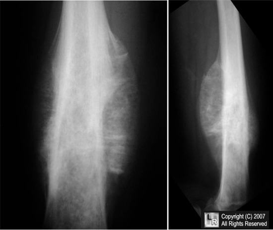
Parosteal Osteosarcoma


Introduction
  • low grade osteosarcoma
  • Age & location
    • more common in females, age 30-40
    • previous exam question ages: 75, 74 yrs
    • occurs on surface of metaphysis of long bones
      • most common sites include posterior distal femur, proximal tibia, and proximal humerus
      • marrow invasion in 25%
Symptoms
  • Presentation
    • often presents as a painless mass
    • can limit joint motion (knee)
Imaging
  • Radiographs
    • heavily ossified, lobulated mass arising from cortex (appears as if sticking to cortex)
  • Bone scan
    • mandatory and always hot
  • CT chest
    • mandatory to rule out pulmonary mets
  • MRI
    • mandatory to determine soft tissue involvement and skip lesions
Histology
  • Characteristic histology
    • regularly arranged normal osseous trabeculae 
    • slightly atypical spindle cells within trabeculae
    • cartilage is often present and may take the form of a cartilage cap
  • Pathologist ocassionally mistakes for fibrous dysplasia
    Treatment
    • Operative
      • wide local surgical excision
        • often curative
        • chemotherapy not indicated unless there is a high grade component
    Prognosis
    • 95% long term survival when local control has been achieved
      • dedifferentiation is a poor prognostic factor
    Groups & Differentials
    • Fibrous dysplasia (similar on histology, but xrays are different)
    • Myositis Ossificans Traumatica (juxtaposed to bone)
    • Osteochondral exostosis (shares cortex with bone)
    • Developmental defect at insertion of adductor magnus