How to use this site?

Please click on the comments to see the right option from the choices given

Dear Medicos,
This site contains a comprehensive list of medical PG entrance questions asked in various PG entrance examination throughout India like AIIMS, AIPGEE, PGI CHANDIGARH, JIPMER, CMC VELLORE .... and various state entrance exams like KERALA, TAMIL NADU, KARNATAKA, DELHI .... and also private entrances like COMEDK, MANIPAL etc...


SEARCH THE WEB

20110921

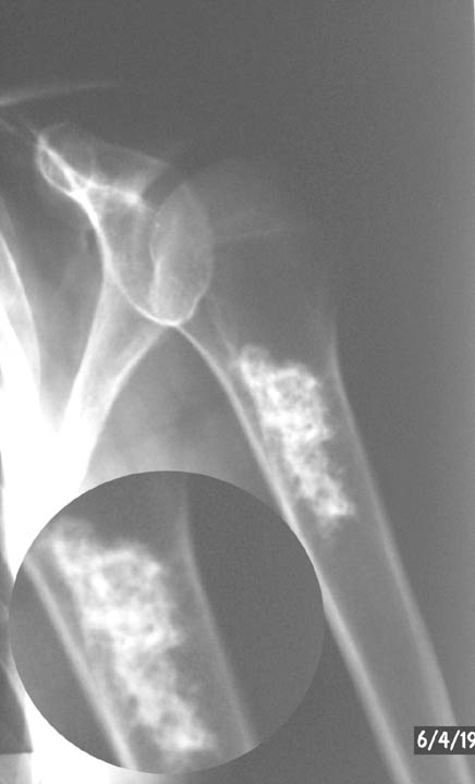
Enchondroma


Introduction
  • A benign chondrogenic lesion caused by an abnormality of chondroblast function in the physis
    • chondroblasts escape the physis and proliferate in metaphysis 
    • 2nd most common benign cartilage lesion (osteochondroma is most common)
  • Age & location
    • age range is 10-60 years
    • occurs in medullary cavity in diaphysis or metaphysis
    • hand is most common location (60%)
      • presentation as pathological fractures common
    • other locations include the femur (20%) and proximal humerus (10%)
  • Include
    • solitary enchondroma
      • 1% chance of malignant transformation (chondrosarcoma)
    • Ollier's disease (multipe enchondromatosis) 
      • no genetic predisposition has been identified
      • multiple enchondromas in long bone diaphysis
      • 10-30% chance of malignant transformation into chondrosarcoma
        • however associated with high rate of both skeletal and non-skeletal malignancies
      • involved bones are dysplastic
    • Maffucci's syndrome
      • no genetic predisposition has been identified
      • multiple enchondromas and soft-tissue angiomas
      • 100% chance of malignant transformation into chondrosarcoma 
      • increased risk of visceral malignancies (astrocytoma, GI malignancy)
Symptoms
  • Presentation
    • most are asymptomatic 
    • often present as pathologic fx of the hand but asymptomatic in foot
  • Physical exam

    • enchondroma may disrupt the growth plate and lead to shortening and angular deformities  
Imaging
  • Radiograph
    • "pop-corn" mineralization (calcification), stippled ringsin diaphyseal or metaphyseal medullary cavity 
    • occasionally may have purly lytic appearance (especially in hand) 
  • Bone scan may be helpful and is usually minimally warm
  • MRI may show steak of cartilage or "sled runner tracks" 
Histology
  • Classic characteristics include
    • hypocellular with bland hyaline cartilage (blue balls of cartilage)   
    • lesions in hand are more hypercellular and even display some worrisome histologic characteristics
    • small chondroid cells in lacunar spaces 
Differential
  • Must differentiate from a bone infarct ("smoke-up-chimney" appearance)
Treatment
  • Nonoperative
    • observation
      • treatment for most enchondromas 
      • watch with serial xrays for interval growth (obtain xrays at 3 months and 1 year from time of presentation)
  • Operative
    • curettage and bone grafting
      • indications
        • lesion that shows any change on serial xrays
        • hand fractures after fracture has first been allowed to heal with imobilization
  • Ollier's disease and Maffucci's
    • treat deformity with diaphysectomy
    • watch lesions carefully for malignant transformation

No comments: