A comprehensive question bank for indian medical PG preparations- AIIMS, ALL INDIA, JIPMER, PGI, state exams etc. Visual and audio content prepared in view of upcoming pattern of NEET (National Eligibility & Entrance Test). Best wishes for your preparation! AIPGE content updated with emphasis on recent questions.
How to use this site?
Please click on the comments to see the right option from the choices given
This site contains a comprehensive list of medical PG entrance questions asked in various PG entrance examination throughout India like AIIMS, AIPGEE, PGI CHANDIGARH, JIPMER, CMC VELLORE .... and various state entrance exams like KERALA, TAMIL NADU, KARNATAKA, DELHI .... and also private entrances like COMEDK, MANIPAL etc...
Pages
SEARCH THE WEB
20150721
Vibrio parahemolyticus is seen in undercooked:
Labels:
Bacteriology,
DNB Dec 09,
Food Poisoning,
Microbiology
20150718
Accumulation of homogentisic acid causes:
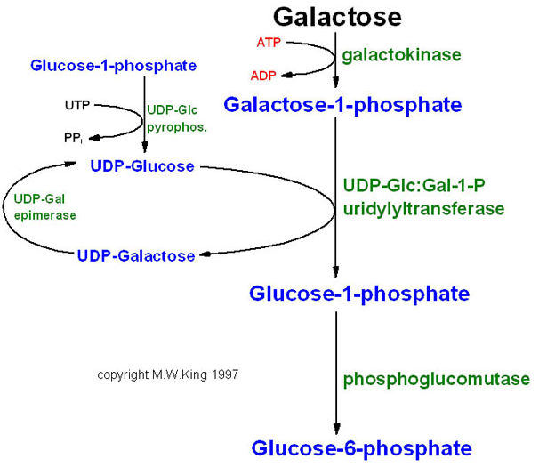
Most common enzyme deficiency responsible for galactosemia is:
a. UDP galactose epimerase
b. Galactokinase
c. Galactosidase
d. Galactose-1-phosphate uridyl transferase
b. Galactokinase
c. Galactosidase
d. Galactose-1-phosphate uridyl transferase

 Types
Types
Galactose is converted into glucose by the action of three enzymes, known as the Leloir pathway. There are diseases associated with deficiencies of each of these three enzymes:
| Type | Locus | Enzyme | Name | |||
| Type 1 | 9p13 | galactose-1-phosphate uridyl transferase | classic galactosemia | |||
| Type 2 | 17q24 | galactokinase | galactokinase deficiency | |||
| Type 3 | 1p36-p35 | UDP galactose epimerase | galactose epimerase deficiency, UDP-Galactose-4-epimerase deficiency |
Ans. d.
20150709
Ethanol is used in ethylene glycol poisoning because it is:
a. Competitive inhibitor of NADPH oxidase.
b. Competitive inhibitor of alcohol dehydrogenase.
c. Competitive inhibitor of aldehyde dehydrogenase.
d. Non- competitive inhibitor of aldehyde dehydrogenase.

Ans. b.
b. Competitive inhibitor of alcohol dehydrogenase.
c. Competitive inhibitor of aldehyde dehydrogenase.
d. Non- competitive inhibitor of aldehyde dehydrogenase.


Ans. b.
20150705
Source of norepinephrine is:
a. Phenylalanine
b. Alanine
c. Tyrosine
d. Tryptophan


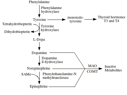


Ans. c. Tyrosine
b. Alanine
c. Tyrosine
d. Tryptophan


Ans. c. Tyrosine
Niemann's pick disease is due to deficiency of:
a. Sphingomyelinase
b. Hexosaminidase - A
c. Aryl sulphatase
d. Galactosidase - A
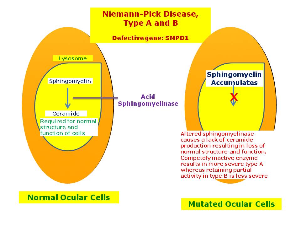
Niemann–Pick disease (/niːmənˈpɪk/ nee-mən-pik)[1] is a group of inherited severe metabolic disorders that allows a certain kind of fat to accumulate in cells. The fat, sphingomyelin, accumulates in lysosomes (membrane-bound organelles in cells). The lysosomes normally transport material through and out of the cell.
Signs and symptoms[edit]
Symptoms are related to the organs in which sphingomyelin accumulates. Enlargement of the liver and spleen (hepatosplenomegaly) may cause reduced appetite, abdominal distension, and pain. Enlargement of the spleen (splenomegaly) may also cause low levels of platelets in the blood (thrombocytopenia)
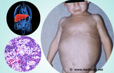

Accumulation of sphingomyelin in the central nervous system (including the cerebellum) results in unsteady gait (ataxia), slurring of speech (dysarthria), and difficulty in swallowing (dysphagia).Basal ganglia dysfunction causes abnormal posturing of the limbs, trunk, and face (dystonia). Upper brainstem disease results in impaired voluntary rapid eye movements (supranuclear gaze palsy). More widespread disease involving the cerebral cortex and subcortical structures causes gradual loss of intellectual abilities, causing dementia and seizures.
Bones can also be affected: symptoms can include enlarged bone marrow cavities, thinned cortical bone, or a distortion of the hip bone called coxa vara. Sleep-related disorders, such as sleep inversion, sleepiness during the day and wakefulness at night, can occur. Gelastic cataplexy, the sudden loss of muscle tone when the patient laughs, is also seen.

Mutations in the SMPD1 gene cause Niemann–Pick disease types A and B. They stop the body from making an enzyme, acid sphingomyelinase, that breaks down lipids.[3]
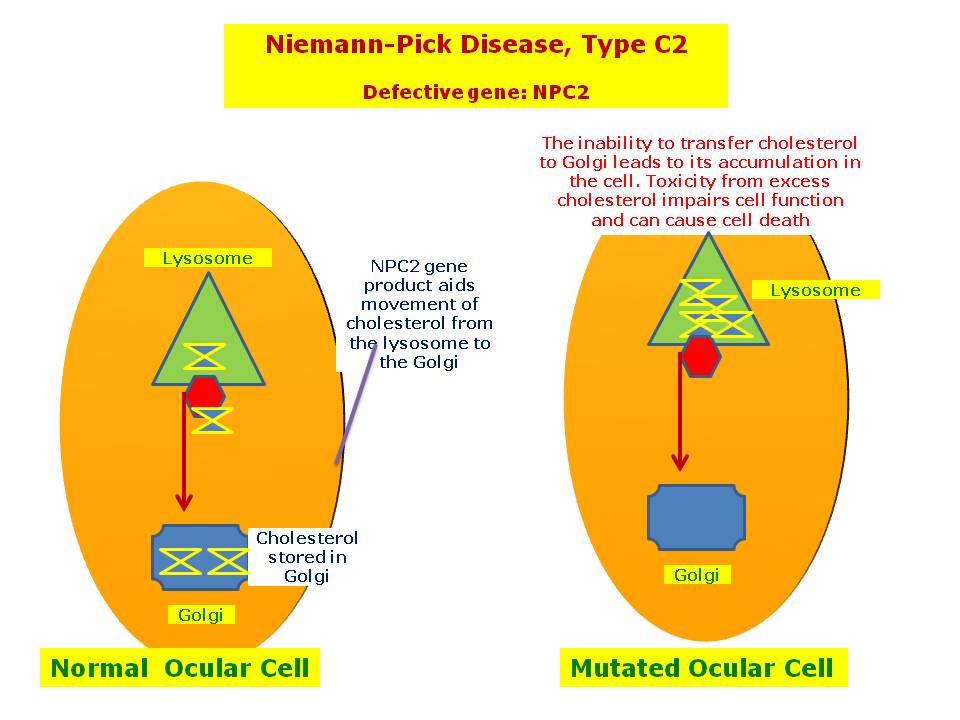
Mutations in NPC1 or NPC2 cause Niemann–Pick disease, type C (NPC), which affects a protein used to transport lipids.
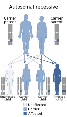
Niemann–Pick disease is inherited in an autosomal recessive pattern, which means both copies, or alleles, of the gene must be defective to cause the disease.

Classification[edit]
- Niemann–Pick disease, SMPD1-associated, which includes types A and B
- Niemann–Pick disease type A: classic infantile
- Niemann–Pick disease type B: visceral
- Niemann–Pick disease, type C: subacute/juvenile, includes types C1 (95% of type C) and C2. Type C is the most common form of the disease[3] Type C2 is a rare form of the disease
Pathophysiology[edit]
Niemann–Pick diseases are a subgroup of lipid storage disorders called sphingolipidoses in which harmful quantities of fatty substances, or lipids, accumulate in the spleen, liver, lungs, bone marrow, and brain.
In the classic infantile type-A variant, a missense mutation causes complete deficiency of sphingomyelinase. Sphingomyelin is a component of cell membrane including the organellar membrane, so the enzyme deficiency blocks degradation of lipid, resulting in the accumulation of sphingomyelin within lysosomes in the macrophage-monocyte phagocyte lineage. Affected cells become enlarged, sometimes up to 90 μm in diameter, secondary to the distention of lysosomes with sphingomyelin and cholesterol. Histology shows lipid-laden macrophages in the marrow and "sea-blue histiocytes" on pathology. Numerous small vacuoles of relatively uniform size are created, giving the cytoplasm a foamy appearance
Ans. a. Sphingomyelinase
Pick's disease, a type of Fronto-temporal Dementia, is a rare neurodegenerative disease that causes progressive destruction of nerve cells in thebrain. Symptoms include dementia and loss of language (aphasia). While some of the symptoms can initially be alleviated, the disease progresses and patients often die within two to ten years.[1] A defining characteristic of the disease is build-up of tau proteins in neurons, accumulating into silver-staining, spherical aggregations known as "Pick bodies"
20150704
Pulse pressure is affected by:
a. Stroke volume
b. Compliance of aorta
c. Ejection fraction
d. All

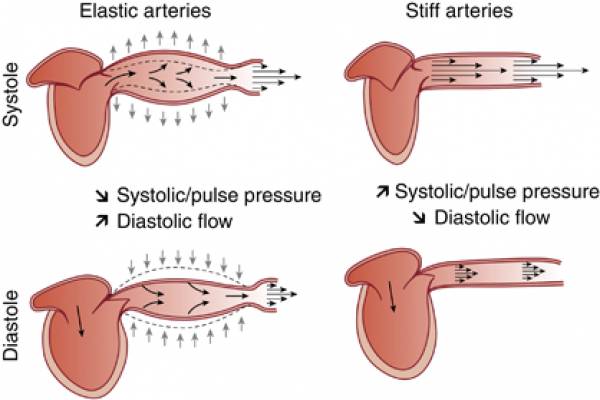
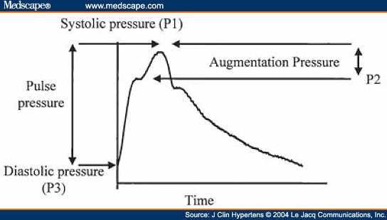
Ans. d. All
b. Compliance of aorta
c. Ejection fraction
d. All


Ans. d. All
Hypoxia without cyanosis is:
a. Stagnant
b. Hypoxic
c. Anemic
d. Histotoxic
Ans c. Anemic
b. Hypoxic
c. Anemic
d. Histotoxic
Ans c. Anemic
Maximum velocity of conduction is seen in:
a. SA node
b. AV node
c. Bundle of His
d. Purkinje fibres

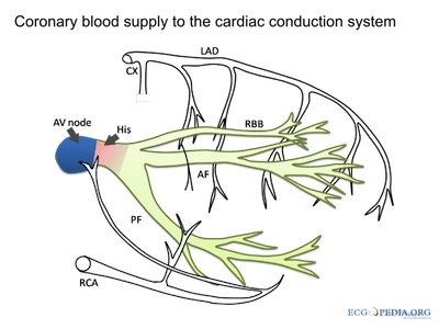
Ans: d. Purkinje
b. AV node
c. Bundle of His
d. Purkinje fibres


Ans: d. Purkinje
Nitric oxide is produced from
a. Endothelium
b. RBC
c. Platelets
d. Lymphocytes

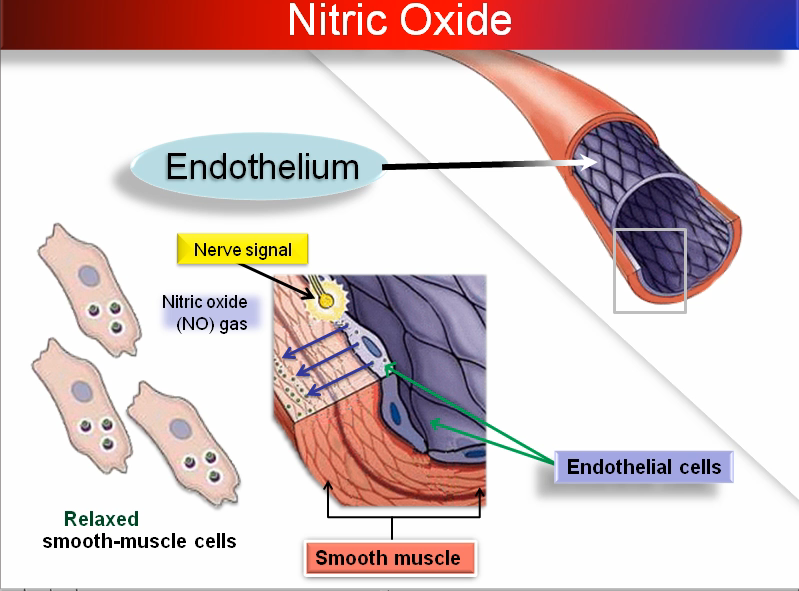
Ans a. Endothelium
b. RBC
c. Platelets
d. Lymphocytes


Ans a. Endothelium
Ion for conversion of prothrombin to thrombin is:
a. Sodium
b. Potassium
c. Magnesium
d. Calcium

Ans : d. Ca
b. Potassium
c. Magnesium
d. Calcium

Ans : d. Ca
Medial geniculate body is related to:
a. Vision
b. Hearing
c. Balance
d. Smell
Ans . B. Hearing
b. Hearing
c. Balance
d. Smell
Ans . B. Hearing
Contractile unit of muscle is:
a. Sarcomere
b. Sarcolemma
c. Myofibril
d. Sarcotubular system

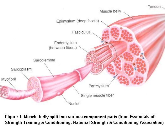

Ans a. Sarcomere
b. Sarcolemma
c. Myofibril
d. Sarcotubular system


Ans a. Sarcomere
Preload measures:
a. End systolic volume
b. End diastolic volume
c. Peripheral resistance
d. Stroke volume

Ans b. EDV
b. End diastolic volume
c. Peripheral resistance
d. Stroke volume

Ans b. EDV
TRH stimulates the release of:
a. Prolactin only
b. TSH only
c. Both
d. None


Ans. c. Both
b. TSH only
c. Both
d. None


Ans. c. Both
Transport of two substances in same direction is called as:
a. Symport
b. Antiport
c. Exocytosis
d. Pinocytosis

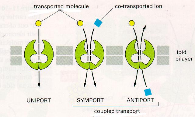
Ans a. Symport
b. Antiport
c. Exocytosis
d. Pinocytosis

Ans a. Symport
Most abundant extracellular ion is:
20150703
All develop from 2nd branchial arch except:
Goblet cells are present in all except?
a. Small intestine
b. Large intestine
c. Esophagus
d. Stomach

Goblet cells: glandular simple columnar epithelial cells.
- secretes mucin, which dissolves in water to form mucus.
- use apocrine and merocrine methods.
- found in epithelial lining of intestinal and respiratory tracts.
: Trachea, bronchus, bronchioles
: Small intestine, Colon
: Conjunctiva in upper eyelid

Clinical significance
Goblet cell carcinoids are a class of rare tumors that form as a result of an excessive proliferation of both goblet and neuroendocrine cells. The majority of these tumors arise in the appendix and may present symptoms similar to the much more common acute appendicitis.The main treatment for localized goblet cells tumors is removal of the appendix, and sometimes removal of the right hemicolon is also performed.Disseminated tumors may require treatment with chemotherapy in addition to surgery.
Goblet cells may be an indication of metaplasia, such as in Barrett's esophagus.
Ans d. Stomach
b. Large intestine
c. Esophagus
d. Stomach

Goblet cells: glandular simple columnar epithelial cells.
- secretes mucin, which dissolves in water to form mucus.
- use apocrine and merocrine methods.
- found in epithelial lining of intestinal and respiratory tracts.
: Trachea, bronchus, bronchioles
: Small intestine, Colon
: Conjunctiva in upper eyelid

Clinical significance
Goblet cell carcinoids are a class of rare tumors that form as a result of an excessive proliferation of both goblet and neuroendocrine cells. The majority of these tumors arise in the appendix and may present symptoms similar to the much more common acute appendicitis.The main treatment for localized goblet cells tumors is removal of the appendix, and sometimes removal of the right hemicolon is also performed.Disseminated tumors may require treatment with chemotherapy in addition to surgery.
Goblet cells may be an indication of metaplasia, such as in Barrett's esophagus.
Ans d. Stomach
Which layer of scalp is vascular?
a. Skin
b. Subcutaneous tissue
c. Aponeurosis
d. Loose connective tissue

Ans. b. Subcutaneous dense connective tissue
b. Subcutaneous tissue
c. Aponeurosis
d. Loose connective tissue

Ans. b. Subcutaneous dense connective tissue
Subscribe to:
Posts (Atom)

