| Symptoms |
- Presentation
- often presents as a painless mass
- can limit joint motion (knee)
|
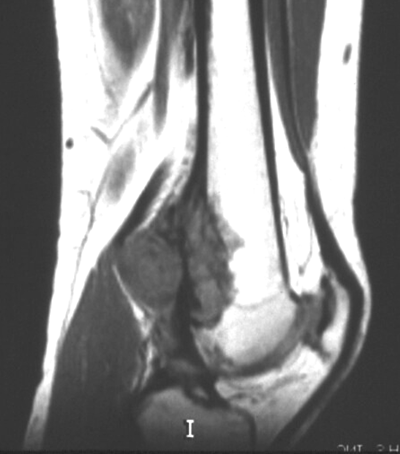
| Imaging |
- Radiographs
- heavily ossified, lobulated mass arising from cortex (appears as if sticking to cortex)
- Bone scan
- CT chest
- mandatory to rule out pulmonary mets
- MRI
- mandatory to determine soft tissue involvement and skip lesions
|
| Histology |
- Characteristic histology
- regularly arranged normal osseous trabeculae
- slightly atypical spindle cells within trabeculae
- cartilage is often present and may take the form of a cartilage cap
- Pathologist ocassionally mistakes for fibrous dysplasia
|
|
|
| Treatment |
- Operative
- wide local surgical excision
- often curative
- chemotherapy not indicated unless there is a high grade component
|
| Prognosis |
- 95% long term survival when local control has been achieved
- dedifferentiation is a poor prognostic factor
|
| Groups & Differentials |
- Fibrous dysplasia (similar on histology, but xrays are different)
- Myositis Ossificans Traumatica (juxtaposed to bone)
- Osteochondral exostosis (shares cortex with bone)
- Developmental defect at insertion of adductor magnus
|


No comments:
Post a Comment